

Quick Links
Web-Based Instruction
Veterinary Neurobiology Courseware
(Arranged Categorically)
To view a web site click its title.
General Courseware Recommendations for Veterinary Students
Recommends courseware available on this Veterinary Anatomy web site for veterinary students studying neurobiology.
Brain Neuroanatomy Courseware:
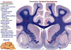
Canine Brain Transections
A web site presenting neuroanatomy of the canine brain as seen in 20 transverse sections labeled to identify what veterinary students are expected to know in a Veterinary Neurobiology course. The web site has two divisions: 1] Brain Transection Levels, which correlates brain levels with brain regions, and 2] Brain Transection Atlas, which identifies anatomical components within brain transections. The atlas features two modes of identification: select a name & see the corresponding structure identified and described, or select a structure & see its name high-lighted. Users can toggle between the two divisions and navigate via an index screen showing small images of all sections. Pop-up glossary statements have been added. When a term is clicked in an Atlas plate, a dot label shows its location and a glossary statement describes it.
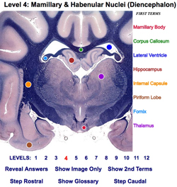
Canine Brain Sections Tutor
This interactive web app is intended to engage student interest and encourage them to study internal brain anatomy at their own pace. Twelve brain transection levels are selectable numerically or via an image index page. Each brain image presents labeled structures and random terms; either may be clicked to reveal associations by color match. At any time a student may view: a preliminary vs advanced term-set, a glossary of term descriptions, the brain image only, or lines connecting all structures and terms. The web app includes a Quiz Mode that presents unlimited random questions (a random level, a random structure, plus five randomly ordered terms). Alternatively, clicking or tapping the structure reveals the answer.
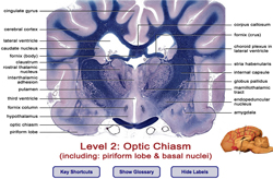
Canine Brain Atlas
This Brain Atlas web site consists of twelve transverse levels through a dog brain. The Atlas is accompanied by a Glossary-Index of brain anatomy terms. Labels may be toggled on/off and levels may be navigated with a mouse or key strokes. (Page width is 1200 pixels; a print version of this Atlas is available in A Practical Guide to Canine & Feline Neurology, 2nd ed. by Curtis W. Dewey, Wiley-Blackwell 2008.)

Brain Gross Anatomy Images
This Brain Gross Anatomy web site presents brain dissection images intended for veterinary students studying neurobiology at the University of Minnesota. Brain components are shown in intact or dissected brains of horse, cow, sheep, dog, or cat. Images of dissected brains are organized by anatomical region and by viewing perspective. Each brain image has an accompanying caption and labels that can be toggled on/off.
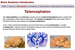
Brain Anatomy Introduction Lab
This web site is organized as the Brain Introduction section of a Neuroanatomy Lab Guide. It was developed to present beginning brain anatomy from different perspectives via a variety of color images of brain dissections and transections. The site is regionally organized corresponding to sections of the printed Lab Guide. Terms that students should learn for this Lab are presented in bold type.
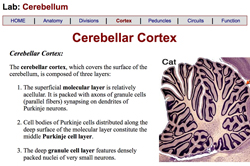
Cerebellum Lab
This web site is organized as the Cerebellum section of a Neuroanatomy Lab Guide. It presents cerebellar anatomy and function via diagrams and images of brain dissections and tissue sections. The site is organized as follows: Anatomy, Cerebellar Divisions, Cortex, Peduncles, Circuits, and Function. Terms that students should learn for this Lab are presented in bold type.
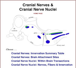
Cranial Nerves & Cranial Nerve Nuclei
This web app presents a stand-alone interactive tutorial for canine cranial nerves and cranial nerve nuclei, including: a cranial nerve innervation summary table; canine brain attachment sites of cranial nerve roots; identification of nuclear neuron columns per fiber-type in brainstem cartoons; cranial nerve nuclei shown within brainstem transverse tisue sections; and cranial nerve & nuclei innervation targets. The web app may be used to supplement neuroanatomy lab materials pertaining to cranial nerves and nuclei.

Cranial Nerve Nuclei - Animated Quizzes
Animated quizzes are provided for students to reinforce their knowledge of cranial nerve nuclei with respect to name, innervation targets, associated cranial nerves, and fiber-type per nucleus. The quizzes facilitate student manipulation of screen features related to dorsal, sagittal and transverse cartoons of cranial nerve nuclei. Correct/incorrect responses are signaled by changes in screen appearance. A comprehensive explanation summary per nucleus is also included.
Brain MRI Courseware:
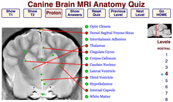
Canine Brain MRI Atlas
This web site presents labeled transverse views of a Beagle Brain obtained by Magnetic Resonance Imaging: T2-weighted, T1-weighted, and Proton-Density-weighted MRIs are included. Three viewing options are presented: 1] MRIs are paired with stained tissue sections to facilitate neuroanatomy identification; 2] brain MRIs are shown in situ within transverse views of the Beagle head; and 3] MRIs are featured in animated interactive quizzes intended to offer knowledge self-assessment of MRI brain anatomy (click/tap a term and then its corresponding target; if correct, a connecting line will appear).
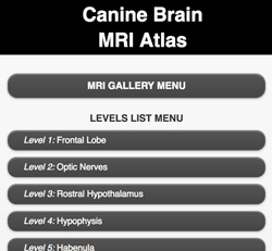
mobile MRI Brain Atlas
This web app presents proton-density-weighted MRI images paired with brain-transection images of a dog brain. The app is intended to provide quick access to canine brain MRI anatomy through a smart phone. Particular brain levels are accessible via a Gallery Menu (tap a small image) or a List Menu (tap a text button). At each level, a button toggles between MRI and brain section images. Labels per image can be switched on/off by tapping the image (or a Show/Hide button). Rostral/caudal buttons are available for navigation to adjacent levels. This web app can be viewed on a smart phone, a tablet, or a computer screen.
Spinal Cord Courseware:
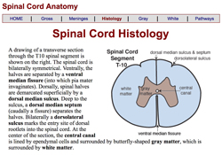
Spinal Cord Anatomy Lab
This web site is organized as the Spinal Cord section of a Neuroanatomy Lab Guide. It presents spinal cord anatomy via color images, including gross anatomy, segment-vertebrae relationships, meninges, blood supply, histology of spinal gray and white matter, and spinal tracts. Terms that students should learn are presented in bold type.
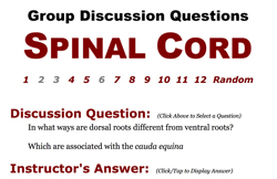
Spinal Cord - Discussion Questions
This web site presents thoughtful questions related to the Spinal Cord. It is intended for group discussion. The Group selects a question at random and attempts to achieve a consensus answer. Then that consensus answer is compared to an instructor's answer.
General Nervous System Courseware:
Interactive Neuroanatomy Quiz
This web site enables students to self-evaluate knowledge of basic neuroanatomy, in an engaging interactive manner. Within a selected topic, individual image screens present six question boxes and six randomly positioned answer boxes. Students either drag a question box to its correct answer box, or sequentially click first a question box and then an answer box. Correct choices are signaled by immediate changes in borders, colors & numbers for correct answers; incorrect answers evoke a mismatch pop up. A poster displaying selected question images is available: here
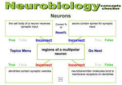
Neurobiology Concepts Checker
This web site focuses on veterinary neurobiology concepts, giving students an opportunity to clarify conceptual understanding via self-assessment. Per subject category, a series of query screens is presented randomly. Each screen consists of a centrally placed statement surrounded by four randomly positioned phrases that are either true or false. A Show Answer button reveals your success and offers a conceptual explanation.
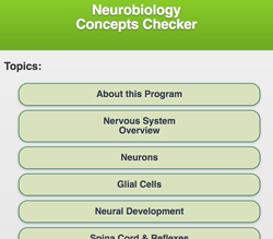
Neurobiology Concepts Checker (mobile)
This Neurobiology Concepts Checker is a web app designed for smart phones. For each selected Neurobiology Topic, pertinent concepts are presented in the form of a conceptual statement and four True/False questions.
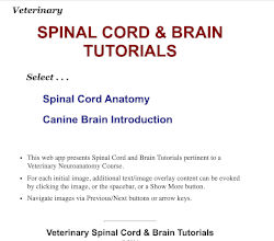
Spinal Cord & Brain Tutorials
This web site presents Spinal Cord & Brain Tutorials corresponding to content taught in an instructional Veterinary Neuroanatomy Lab. The Tutorials can be used to complement or substitute for a Lab Manual. Each tutorial page displays an initial image; additional text and/or images are revealed by clicking the image or the spacebar or a Show More button. Images are navigated via Previous/Next buttons or arrow keys.
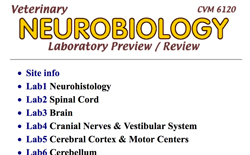
Neurobiology Labs Overview — Preview & Review Images
This is the initial web site developed for self-study of Neuroanatomy Lab subject matter by veterinary students at the University of Minnesota (the site is gradually being supplanted by individual Lab web sites). Lab content, arranged for each of eight Neuroanatomy Labs, is presented in the form of images with captions, including links to other images.
Peripheral Nervous System Courseware:
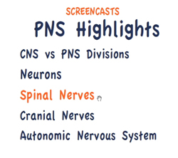
PNS Highlights Video Screencasts
Narrated synopses (screencasts) highlighting the carnivore Peripheral Nervous System are presented as streaming video clips (800 x 600px .mp4). The video topics are: PNS vs CNS divisions of the nervous system; Neuron and Neurohistology background information; Spinal Nerves; Cranial Nerves; and Autonomic Nervous System.

Cranial Nerves - Animated Quizzes
Animated quizzes are presented, enabling students to reinforce their knowledge of names/numbers, innervation targets, and fiber-type content of cranial nerves. The quizzes reveal correct answers in response to drag or click/tap operations by students. Clicking RESET generates a new randomized version for each quiz. A summary page of cranial nerve fiber-types is included.
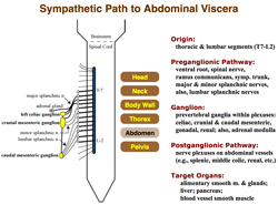
Autonomic Nervous System
This web site presents autonomic nervous system anatomy and autonomic physiology/pharmacology. Visceral efferent nerve pathways to canine body regions are emphasized. The web site features animated tutorials for learning pathways and animation quizzes for tracing one's knowledge of sympathetic and parasympathetic pathways to regional visceral organs.
Neurohistology Courseware:
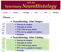
Neurohistology ATLAS
This site presents an image Atlas, consisting of over 60 labeled images with captions. Atlas images are linked circularly by Previous/Next buttons. To facilitate Atlas navigation, an associated Image Catalog features image buttons that are reciprocally linked to Atlas images. Web site images are grouped into five neurohistology categories: Neuron & Synapse; Neuroglia & Myelin; Central Nervous System; Peripheral Nervous System; and Meninges.
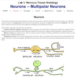
Neurohistology Lab
This web site reproduces the contents of a printed Neuroanatomy Lab Guide, presenting images in color. Divisions of the Lab Guide include: neurons, neuroglial cells, peripheral nerves, spinal meninges, spinal ganglia and lamellar corpuscle receptors. Neurohistology Lab objectives are included.
Neuroembryology Courseware:
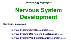
Embryology Highlights: Nervous System Development
This web site presents links to narrated synopses (screencasts) of nervous system embryonic development. Topics include: Early neural development, CNS development, and PNS & Meninges development. The screencasts were produced with Camtasia software; they are presented as streaming video clips (800 x 600px .mp4).
Clinical Related Courseware:
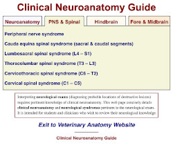
Clinical Neuroanatomy Guide
Interpreting neurological exams (i.e., diagnosing probable locations of destructive lesions) requires pertinent knowledge of clinical neuroanatomy. This web app concisely details clinical neuroanatomy and neurological syndromes pertinent to the neurological exam. All of the content is immdediately available within a single page by clicking tabs and accordian elements that are plainly visible as Contents links. The web app is is intended for students and clinicians who wish to review their neurological knowledge.
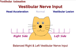
Vestibular Animations
Animations depict balanced vestibular nerve input and vestibular nerve imbalances produced by either head acceleration or destructive lesions. Explanations of vestibular reflexes and the clinical vestibular syndrome are included as background information.
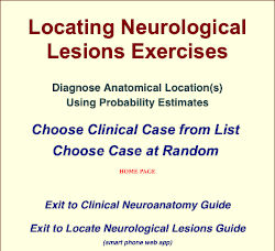
Locating Neurological Lesions Exercises
This web app provides opportunities to exercise neuroanatomy knowledge to localize destructive lesions based on clinical signs, for a dozen clinical cases. Intended for veterinary students who have had or are studying neuroanatomy, the web app promotes a probability approach for diagnosing lesion location. Via buttons, you may view: patient information (an image and neurological exam narrative); instructor entries (probability opinions of an experienced instructor); lesion location (the actual location(s) determined by necropsy or otherwise); next case (exit to the Home Page for a next case list or random case selection).
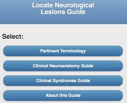
Locate Neurological Lesions Guide
This web app presents Clinical Neuroanatomy and Clinical Syndromes Guides. It is intended for veterinary students preparing to diagnose locations of neurological lesions based on clinical signs, using their knowledge of neuroanatomy. The web app was designed for smart phones. Neuroanatomy images may be tapped/clicked to toggle (show/hide) labels.

Video-clips of Clinical Neurology Cases
Intended to enhance neurology instruction, this web site delivers video-clips (640 x 480 .mp4) of selected patients of the University of Minnesota Veterinary Medical Center. The patients exhibited either neurology disorders or normal responses during a clinical neurology exam. The video-clips are organized chronologically, by species, and by syndrome.
- Courseware Recommendations for Veterinary Students
- Canine Brain Transections
- Canine Brain Sections Tutor
- Canine Brain Atlas
- Brain Gross Anatomy Images
- Brain Anatomy Introduction Lab
- Cerebellum Lab
- Cranial Nerves & Cranial Nerve Nuclei
- Cranial Nerve Nuclei - Animated Quizzes
- Canine Brain MRI Atlas
- mobile MRI Brain Atlas
- Spinal Cord Anatomy Lab
- Spinal Cord - Discussion Questions
- Interactive Neuroanatomy Quiz
- Neurobiology Concepts Checker
- Neurobiology Concepts Checker (mobile)
- Spinal Cord & Brain Tutorials
- Neurobiology Labs Overview — Preview & Review Images
- PNS Highlights Video Screencasts
- Cranial Nerves - Animated Quizzes
- Autonomic Nervous System
- Neurohistology ATLAS
- Neurohistology Lab
- Embryology Highlights: Nervous System Development
- Developmental Anatomy: Nervous System & Special Senses
- Clinical Neuroanatomy Guide
- Vestibular Animations
- Locating Neurological Lesions Exercises
- Locate Neurological Lesions Guide (mobile web app)
- Video-clips of Clinical Neurology Cases
- Neurobiology Learning Objects
- Veterinary Neuroanatomy Laboratory Guide (PDF)