Basal Nuclei
CLOSE

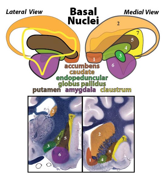
Figure 18—44. Top: Schematic drawing of telencephalic basal nuclei of the right cerebral hemisphere, shown in lateral and medial views. Only the outline of the claustrum is shown in the lateral view. Nuclei names are displayed in colors that match their respective nuclear colors. Names and numbers are listed in approximate medial to lateral order. (Nuclear topography is based on Vakolyuk, N. I. 1974. A Stereotaxic Atlas of Subcortical Nuclei of the Dog’s Brain. Kiev, Academy of Sciences of the Ukrainian SSR.)
Bottom: Approximate transverse sections through the right cerebral hemisphere at the level of the optic chiasm (left) and the frontal lobe (right). Respective basal nuclei are numbered and shown in colors that correspond to those displayed above. (1 = accumbens; 2 = caudate; 3 = endopeduncular; 4 = globus pallidus; 5 = putamen; 6 = amygdala; 7 = claustrum)
Go Top