| PLANE | REGION | Head & Neck | Thorax | Abdomen & Pelvis |
| Sagittal Plane: |
View
Index
|
View
Index
|
View
Index
|
| Transverse Plane: |
View
Index
|
View
Index
|
View
Index
|
| Dorsal Plane: |
View
Index
|
View
Index
|
View
Index
|
|
This web app presents canine planar anatomy via 900 x 600 pixel images of cadaver slabs, obtained by frozen cadaver bandsaw cuts. The head/neck, thorax & abdomen/pelvis are shown in three planes: sagittal [left to right], transverse caudal views [rostral/cranial to caudal], and dorsal [dorsal to ventral] views. At the right, from top to bottom, see examples of a sagittal cadaver slab, a transverse slab, and a dorsal plane slab from three dog heads. (The brain is evident centrally in each image.) A sagittal plane divides the cadaver into right & left parts. A transverse plane divides the cadaver into rostral/cranial & caudal parts. A dorsal plane is cut parallel to the back, dividing the cadaver into top (dorsal) & bottom (ventral) parts. The images displayed in this web site were obtained in the process of authoring: Feeney D.A., Fletcher, T. F., and Hardy, R. M. 1991 Correlative Imaging Anatomy of the Normal Dog. Ultrasound and Computed Tomography. W.B. Saunders Co., Philadephia. Navigation Note: Keyboard arrow keys may be used to navigate along an image series. Use the spacebar or click/tap an image to toggle labels. |
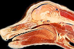 Sagittal Plane
|
|
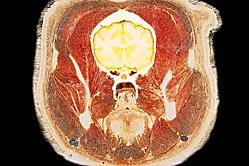 Transverse Plane
|
||
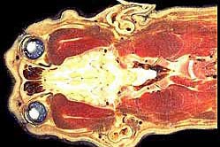 Dorsal Plane
|
© 2017
T.F. Fletcher fletc003@umn.edu
R.M. Hardy hardy001@umn.edu
D.A. Feeney feene001@umn.edu
Last modified April 2021
Supported by University of Minnesota College of Veterinary Medicine
The University of Minnesota is an equal opportunity educator and employer.