
Lab 2 - Image 2
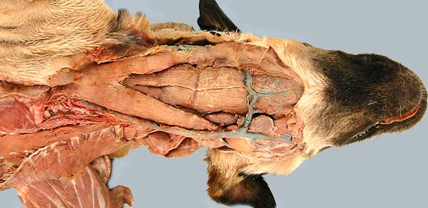
Both sides of the neck are dissected in this ventral view, but you should dissect only the left side. The sternocephalicus m. (1) originates from the manubrium of the sternum (right and left muscles are fused initially). The sternohyoideus m. (2) runs from the sternum to the basihyoid bone. Right and left muscles are separated by a raphe (seam). The sternothyroideus m. (3) is positioned lateral to the sternohyoideus m.
You should not dissect the head at this time. In this specimen, head dissection shows branches of the external jugular vein, including an anastomosis (connection) between right and left sides.