
LAB 2 Introduction
Ventral Neck Muscles and
Extrinsic Muscles of the Thoracic Limb
(Guide to the Dissection of the Dog, 8th ed., pp. 20-26)
CONTENTS:
Lab Objectives:
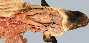
• Identify three ventral neck mm. that run from the sternum to the skull, or hyoid bone, or larynx
(sternocephalicus m., sternohyoideus m., and sternothyroideus m.).
• Notice the carotid sheath (cervical deep fascia surrounding the common carotid a., etc.).
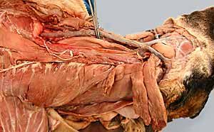
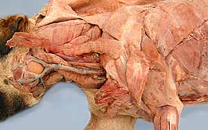
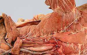
• Examine and transect (the remaining) five of the eight extrinsic muscles of the thoracic limb:
- omotransversarius m. (covering the superficial cervical lymph node)
- trapezius m. (has cervical and thoracic parts)
- rhomboideus m., three divisions: r. capitis, r. cervicis, and r. thoracis
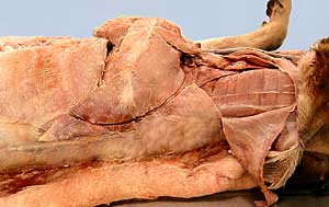
- latissimus dorsi m. (aponeurosis from thoracolumbar deep fascia)
- serratus ventralis m. (has cervical and thoracic parts)
• Detach the thoracic limb from the cadaver.
Anatomical Terms:
Ventral Neck Muscles and Cervical Deep Fascia
sternocephalicus m. (mastoid & occipital parts)
sternohyoideus m.
sternothyroideus m.
superficial cervical lymph node [palpate]
deep fascia of the neck
carotid sheath
median raphe (dorsal)
trachea
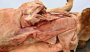
Thoracic Limb Extrinsic Muscles (continued)
omotransversarius m. (covering the superficial cervical lymph node)
trapezius m. (cervical & thoracic parts) [palpate]
rhomboideus m. [palpate] (capitis, cervicis & thoracis parts)
latissimus dorsi m. [palpate]
(aponeurosis of origin from thoracolumbar deep fascia)
serratus ventralis m. (cervicis and thoracis parts)
Note:
cephal = Greek, head
omo = Greek, shoulder
—oid = Greek, resemblance to —
latissimus = Latin, most wide (L. latus = wide; L. —issimus = most)
Instructor Commentary:
The ventral neck region is clinically significant because it is the site for performing an emergency tracheotomy (to re-establish air flow in cases of laryngeal or pharyngeal obstruction). Find the midline separation between right and left sternohyoideus mm. to reach the trachea.
Extrinsic muscles of the thoracic limb have one attachment on the limb and another attachment on the trunk or neck/head. The extrinsic muscles have two major functions. First, when limbs are bearing weight, they move the trunk and, in particular, the serratus ventralis m. carries the weight of the trunk, since the thoracic limb does not articulate with the thorax. Second, during locomotion in cursorial (running) animals they stretch and contract in controlling movement of the proximal limb (scapula, shoulder, and brachium).
All of extrinsic limb muscles must be transected and nerves & vessels running between the trunk and limb must be cut in order to detach the thoracic limb. The thoracic limb is removed to facilitate its subsequent dissection.
Deep fascia is named in locations where is prominent. The accumulation of cervical deep fascia surrounding the common carotid a. is called the carotid sheath (the term "sheath" is disappointing because the fascia is not distinctly organized). The "sheath" envelops the following structures: common carotid a., vagosympathetic nerve trunk (bound to the artery), internal jugular v., and tracheal lymphatics (which may be collapsed and not evident).
In the absence of bone, corresponding muscles of the right and left sides unite with one another along a fascial seam (raphe). You can see a median raphe along the dorsal neck. The linea alba (white line), the raphe of the ventral abdomen, will be seen later. Also, do not bother to look for supraspinous and nuchal ligaments at this time. They will be seen later in the dissection.
Dissection Steps:
Click to view a PDF list of dissection procedures for this lab:
Show List of Dissection Steps (PDF)
Dissection Images:
Note: Click an image to see it enlarged, view its caption, and toggle its labels.
| 1 | 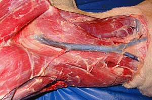 |
 |
2 |
| 3 | 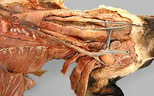 |
 |
4 |
| 5 | 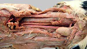 |
 |
6 |
| 7 | 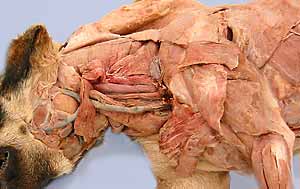 |
 |
8 |
| 9 | 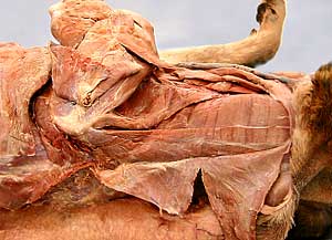 |
 |
10 |
| 11 | 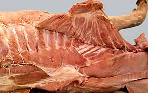 |
 |
12 |