LAB 10 Introduction
Thorax:
Wall and Cavity
(Guide to the Dissection of the Dog, 8th ed., pp. 98-107)
CONTENTS:
Lab Objectives:
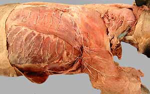
• Within superficial fascia covering the thorax, observe:
- row of dorsal cutaneous nerves
- row of lateral cutaneous nerves
- lateral thoracic n. innervating cutaneous trunci m.
Prepare to Open the Thoracic Cavity
• Transect pectoral muscles on the right side, open the axilla and find:
- lateral thoracic n. innervating cutaneous trunci m.
- lateral thoracic vessels & axillary lymph node
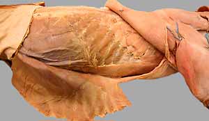
• Reflect muscles from the thoracic wall:
- incise latissimus dorsi (aponeurosis) and reflect it cranially
- incise external abdominal oblique m. and reflect it caudally
- incise rectus abdominis aponeurosis and reflect it caudally
(look for cranial epigastric vessels)
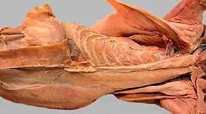
• Open the thoracic cavity bilaterally:
- reflect the sternum (after two bilateral incisions & one caudal incision)
- free the caudal thoracic wall by cutting along the costal arch
- cut ribs dorsally, along the medial edge of the iliocostalis m.
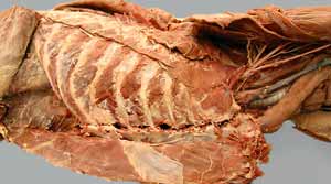
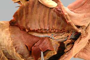
• Identify thoracic cavity contents:
- transversus thoracis m.
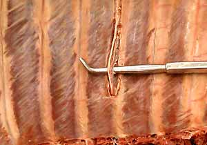
- intercostal vessels and nerves
- internal thoracic vessels (& musculophrenic branch)
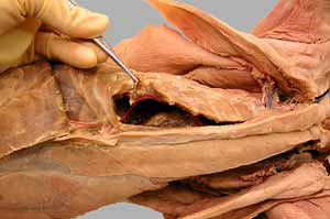
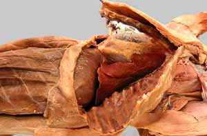
- mediastinum: heart, thymus, etc.
- pleural cavity and pleura:
-- visceral pleura (pulmonary pleura)
-- connecting pleura: pulmonary ligament
-- parietal pleura:
costal pleura
diaphragmatic pleura
mediastinal pleura
plica vena cava
pericardial ligaments
Anatomical Terms:
Thoracic wall structures:
dorsal & ventral intercostal aa.
intercostal nn.
cranial & caudal thoracic mammae
axilla
axillary lymph node
lateral thoracic artery, vein, nerve
cranial epigastric artery (branch of internal thoracic artery)
cranial superficial epigastric artery
transversus thoracis m.
Thoracic cavity, including bilateral pleural cavities:
pleura:
pulmonary (visceral) pleura
parietal pleura:
costal parietal pleura
mediastinal parietal pleura
pericardial mediastinal pleura
plica venae cavae
diaphragmatic parietal pleura
pulmonary ligament (connecting pleura caudal to the root of lung)
mediastinum (region between pleural cavities):
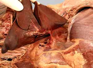
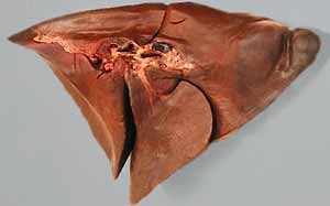
thymus (bilobed organ)
internal thoracic artery (right & left)
musculophrenic artery
Note:
The term pericardium is overloaded with meanings. As a noun it refers to either the
serous membrane or the dense endothoracic fascia surrounding the heart. As an adjective
(pericardial), it refers to the region around the heart.
Instructor Commentary:
The volume of the thymus is age related. It is large in young animals and then gradually becomes smaller as animals age. Its size is related to its immune function.
At the caudal end of the sternum, each internal thoracic vessel terminates in two branches: The cranial epigastic a. supplies rectus abdominis m. and gives off a superficial cranial epigastric a. to the skin. The other branch, the musculophrenic a., consists of multiple small branches in the dog.
The vascular system is arranged in loops designed to maintain blood under pressure for smaller arteries to tap into. The arterial loops are not obvious in muscles, but intercostal arteries can be seen as "loops" between the aorta and bilateral internal thoracic aa. (Fig. 3-5). The rationale for subdividing the intercostals arteries into dorsal and ventral parts relates to the arterial bifurcation ventrally (Fig. 3-5).
Dissection Steps:
Click to view a PDF list of dissection procedures for this lab:
Show List of Dissection Steps (PDF)