
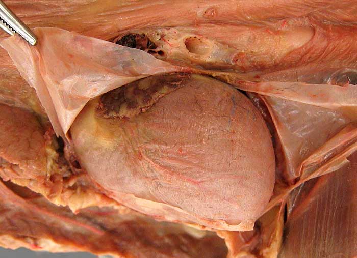
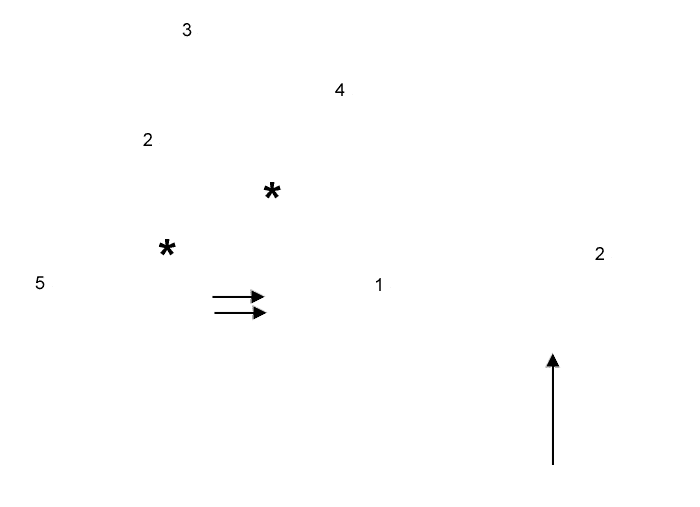
Cat, pericardial sac dissection. The left ventricle of the heart (1) has been exposed by cutting and reflecting the wall (2) of the pericardial sac/cavity (arrow). The left asterisk marks the conus arteriosus region of the right ventricle; the left auricle is marked by the right asterisk. Double arrows indicate the paraconal interventricular groove, the boundary between right and left ventricles.
The surface of the heart is covered by visceral pericardium (epicardium). Parietal pericardium (2) lines the inner surface of the wall of the pericardial sac (the wall is held by forceps). Fibrous pericardium forms the substance of the wall and mediastinal pleural (3) coats the outer surface of the wall. Also notice: root of the left lung (4) and the thymus (5).