
Lab 13 - Image 3
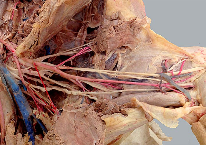
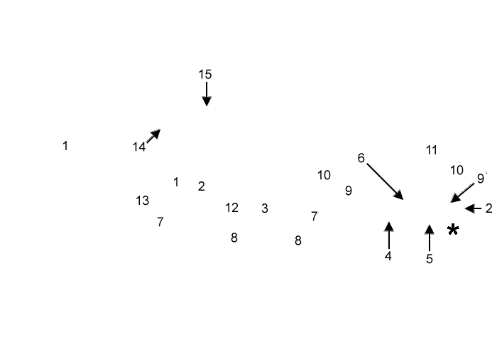
Canine brachial a.: The axillary a. (1) continues as the brachial a. (2). Visible branches of the brachial a. are: deep brachial a. (3), bicipital a. (4), superficial brachial (5), and collateral ulnar a. (6). Veins are poorly injected, but the median cubital v. (asterisk) is evident.
The musculocutaneous n. (7) innervates the biceps brachii m. (8). The median n. (9) runs with the brachial a. The ulnar n. (10) gives off a caudal cutaneous antebrachial n. (11). The large radial n. (12) courses laterally, as does the axillary n. (13). The thoracodorsal n. (14) and the lateral thoracic n. (14) are evident. The latter runs with the lateral thoracic a. and innervates the cutaneus trunci m. (panniculus reflex).