
Lab 14 - Image 4
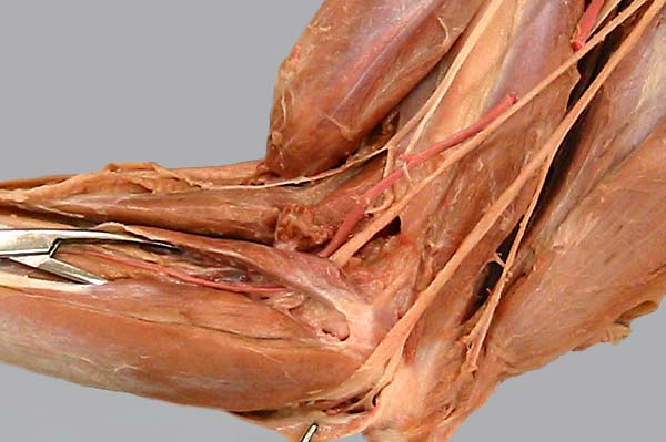
Medial view of the elbow region, the biceps brachii m. has been cut and reflected (asterisks). The brachial a. (1) and median n. (2) pass deep to the pronator teres m. (3). The nerve proceeds deep to the flexor carpi radialis m. (4) as it innervates caudal antebrachial muscles. The ulnar n. (5) also innervates caudal antebrachial muscles. Notice the medial cutaneous antebrachial n. (6) and the caudal cutaneous antebrachial n. (7).