
Lab 14 - Image 6
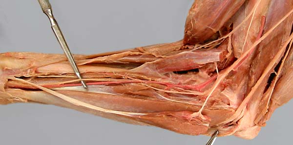
Medial view of the antebrachium. The pronator teres m. (1) and the flexor carpi radialis m. (2) have been cut and reflected. The brachial a. (3) and median n. (4) run together into the proximal antebrachium. After giving off a common interosseus a. (not clearly visible), the brachial a. continues as the median a. (5) which gives off the deep antebrachial a. (6). The median n. (elevated by a probe) runs with the median a. toward the carpal canal.