
LAB 14 Introduction
Thoracic Limb:
Distal Vessels and Nerves
(Guide to the Dissection of the Dog, 8th ed., pp. 139-147)
CONTENTS:
Lab Objectives:
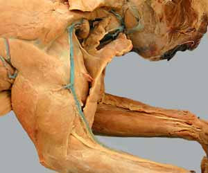
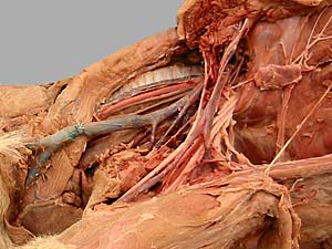
• Continue identifying nerves in the brachium:
- radial n. (runs from medial to lateral on the brachialis m.)
- median n. (runs with the brachial a. on the medial side of the brachium)
- ulnar n. (located caudally on medial side of brachium)
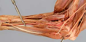
• Remove skin from the antebrachium and manus (you only need to skin the third digit).
Attempt to leave superficial vessels and nerves intact.
• Trace superficial veins of the thoracic limb. In particular, identify the cephalic v. and the median cubital v.
(Notice the nerves & arteries flanking the cephalic v. bilaterally on the cranial antebrachium.)
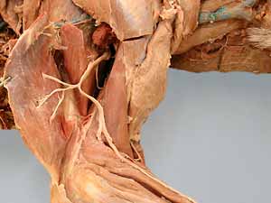
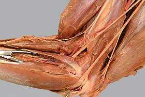
• Incise antebrachal deep fascia and transect pronator teres and flexor carpi radialis mm. to locate brachial a. branches:
- common interosseous a.
- caudal interosseous a. (branch of common interosseous)
- median a.
- deep antebrachial a.
Anatomical Terms:
Thoracic limb: vessels (continued)
cephalic v.
accessory cephalic v.
median cubital v.
axillobrachial v.
omobrachial v.
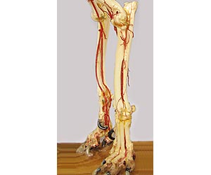
Forearm and Paw:
brachial a. (continued)
common interosseous a. (absent in the cat)
ulnar a.
caudal interosseous a.
cranial interosseous a.
median a. (small in the cat)
deep antebrachial a.
superficial palmar arch
palmar common digital aa. (do not need to identify)
radial a. (large in cat)
Thoracic limb: nerves (continued)
Antebrachium & Manus:
radial n.
deep & superficial branches
median n.
ulnar n.
caudal cutaneous antebrachial n. (do not need to identify)
dorsal branch
palmar branch
Note:
cubitus [Latin] = elbow
Instructor Commentary:
The limbs have two sets of veins. Deep veins are satellites of arteries, and they are named for their corresponding arteries. Superficial veins occupy a subcutaneous location (where they are accessible for intravenous injection or sampling blood). Superficial veins are generally unassociated with arteries. Superficial veins are named "cephalic" in the thoracic limb and "saphenous" in the pelvic limb. A similar arrangement exists in the neck, where the external jugular v. is the superficial vein.
Limb veins have valves about every cm or so. Pressure generated by muscle activity (or external force) moves blood within veins. Because of valves, blood moves only in a proximal direction. Each valve supports its own small column of blood relieving distal veins from having to support a large column of blood. When valves fail their blood columns must be supported distally which tends to cause distal vein enlargement and valve failure. This process is often progressive and results in varicose veins (a human disease).
Digits are numbered from medial to lateral. The pollex (thumb) or the hallux (big toe) are number one. The number identification is retained even when digits are absent. Thus, a dog without a dewclaw has digits 2 through 5; a horse has only digit 3.
You can read about the organization of vessels and nerves in the manus (paw) on page 136 in Guide to the Dissection of the Dog.
(Note: Nomina Anatomica Veterinaria lists the deep antebrachial a. as a branch of the brachial a. rather than the median a., as in the Guide to the Dissection of the Dog.)
Dissection Steps:
Click to view a PDF list of dissection procedures for this lab:
Show List of Dissection Steps (PDF)
Dissection Images:
Note: Click an image to see it enlarged, view its caption, and toggle its labels.
| 1 | 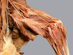 |
 |
2 |
| 3 | 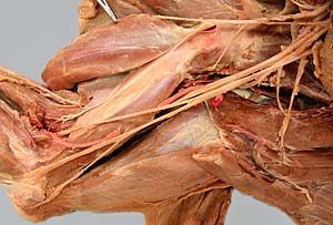 |
 |
4 |
| 5 | 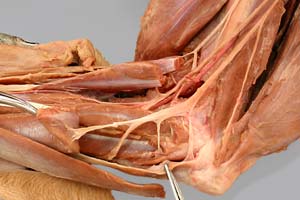 |
 |
6 |
| 7 | 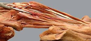 |
 |
8 |
| 9 |  |
 |
10 |
| 11 | 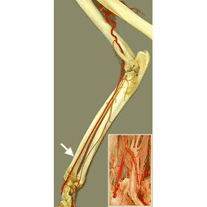 |
 |
12 |
| 13 | 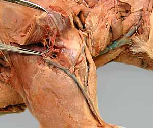 |
 |
14 |