
LAB 15 Introduction
Abdominal Wall & Inguinal Structures
(Vessels and Nerves)
(Guide to the Dissection of the Dog, 8th ed., pp. 148-154)
CONTENTS:
Lab Objectives:
• Reflect skin from the caudal half of the right side and look for superficial inguinal lymph nodes
and cutaneous vessels:
- caudal superficial epigastric a. (from the external pudendal a.)
- look for superficial arterial branches from the four quadrant abdominal blood supply
• Examine ventral branches of lumbar spinal nerves and their lateral cutaneous nn.,
especially the lateral cutaneous femoral n. (last lateral cutaneous nerve).
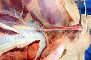
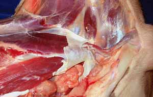
• Identify the inguinal canal and its contents:
- external pudendal vesels and genitofemoral n.
- vaginal process (female) or spermatic cord (male)
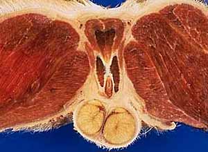
• In the male, examine: testis, epididymis, spermatic fascia (external & internal),
cremaster m. (dog), and components of the spermatic cord:
- vaginal process (parietal, visceral, & connecting tunics)
- ductus deferens and testicular vessels
Anatomical Terms:
Abdominal wall: ventral & lateral
external pudendal a.
caudal superficial epigastric artery
cranial labial/scrotal a.
superficial inguinal lymph nodes
cranial abdominal a. (not usually evident)
deep circumflex iliac a. & v. (will be seen in Lab 18)
lumbar spinal nn. (ventral branches)
cranial iliohypogastric n. (L1 ventral branch)
caudal iliohypogastric n. (L2 ventral branch)
ilioinguinal n. (L3 ventral branch)
lateral cutaneous femoral n. (from L4 ventral branch)
Inguinal Structures
inguinal canal
external pudendal a. & v.
genitofemoral nerve
spermatic cord (male) or vaginal process (female)
MALE
spermatic fascia (loose external & dense internal)
cremaster muscle (dog; usually absent in cats, which have a levator scroti m.)
spermatic cord (begins at the vaginal ring = strict anatomical definition)
vaginal process
parietal & visceral vaginal tunics
mesorchium
mesoductus deferens
ductus deferens
deferent artery/vein
testicular artery/vein
pampiniform (venous) plexus
testis [palpate]
epididymis [palpate] (head, body, tail)
ligament of the tail of the epididymis
proper ligament of the testis
scrotum [palpate]
FEMALE
vaginal process (contains fat & the round ligament of the uterus)
Note:
vaginal [Latin] = sheath
Instructor Commentary:
The scrotum is always located between the anal canal and the penis (in the prepuce). In the cat, the penis is directed caudally and the scrotum is positioned just below the anal canal.
The dog (and most male mammals) have a cremaster muscle to bring the testis close to the body wall. Most cats lack a cremaster m. (it would have to curve to reach the testis); instead, cats have a levator scroti m. that originates from the external anal sphicter, attaches to the scrotal septum, and pulls the entire scrotum closer to the body.
Spermatic fascia surrounds the spermatic cord and lines the scrotum. It is derived from transversalis and subcutaneous fascia of the abdomen and it envelops the parietal tunic of the vaginal process. Spermatic fascia is often divided into internal and external components. Internal spermatic fascia forms a strong membrane (fibrous tunic) to which the cremaster muscle attaches and which is internally lined by parietal vaginal tunic. External spermatic fascia is a loose areolar tissue that surrounds internal spermaltic fascia and cremaster muscle.
Commonly, people use the term spermatic cord to refer to the entire bundle of structures passing between the body wall and the scrotum. However, according to the strict anatomical definition, "spematic cord" pertains only to components within the vaginal cavity (the spematic cord begins at the vaginal ring). Anatomically, "spermatic cord" consists of testicular vessels, the ductus deferens and its vessels and visceral and connecting vaginal tunics, including mesoductus deferens and mesorchium. Thus "spermatic cord" is surounded by parietal vaginal tunic, spermatic fascias and cremaster muscle.
The testicular artery snakes back and forth and testicular veins undergo profuse branching (pampiniform venous plexus). This creates an extensive area of arteriovenous contact for heat conservation. Arterial heat is gradually transferred to returning venous blood with two big benefits: The testes remain cool (the reason they are subcutaneous in the first place) and body heat is conserved (the majority of food calories are required to maintain body heat).
In cervical and thoracic regions, ventral branches of spinal nerves are identified by number, e.g., C-3; T-6; etc. In the lumbar region you can identify the first three ventral branches by number or by name:
L1 = cranial iliohypogastric n.
L2 = caudal iliohypogastric n.
L3 = ilioinguinal n.
Beginning with L-4, spinal nerve ventral branches contribute to the lumbosacral plexus. However, the L-4 ventral branch does give rise to the last of the series of lateral cutaneous nerves. The cutaneous nerve is larger than other and it is named "lateral cutaneous femoral nerve".
Dissection Steps:
Click to view a PDF list of dissection procedures for this lab:
Show List of Dissection Steps (PDF)
Dissection Images:
Note: Click an image to see it enlarged, view its caption, and toggle its labels.
| 1 |  |
 |
2 |
| 3 |  |
 |
4 |
| 5 | 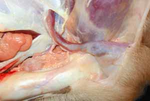 |
 |
6 |
| 7 | 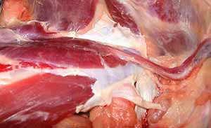 |
 |
8 |
| 9 | 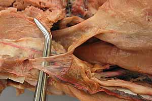 |
 |
10 |