
Lab 20 - Image 14
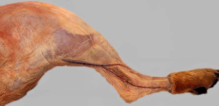
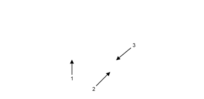
Lateral view of a dog pelvic limb showing the lateral saphenous vein (1). The vein is formed by the union of caudal (2) and cranial (3) branches. The cranial branch of the lateral saphenous vein is generally quite prominent in dogs, but its mobility presents a challenge for venipuncture.