
LAB 22 Introduction
Superficial Structures of the Head
and the Oral Cavity & Pharynx
(Guide to the Dissection of the Dog, 8th ed., pp. 236-248)
CONTENTS:
Lab Objectives:
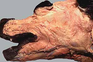
•Dissect muscles of facial expression on the left half of head:
- cutaneous muscles, e.g., platysma muscle
- muscles in walls of lips, cheeks, and eyelids
- superficial muscles that move ear and nose & upper lip
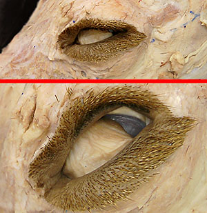
• Examine eyelid features and the conjunctival sac.
• Examine oral cavity, tongue, and salivary glands.
• Identify regions of the pharynx:
- oropharynx (contains palatine tonsil)
- nasopharynx (contains auditory tube opening)
- laryngopharynx
• Optionally, examine osseous features of the nasal cavity.
Anatomical Terms:
Superficial Structures of the Head
philtrum [palpate]
platysma m.
orbicularis oris m. (need not identify)
buccinator m.
levator nasolabialis m.
superior & inferior palpebrae
palpebral fissure
medial & lateral palpebral commissures [palpate]
orbicularis oculi m.
retractor anguli oculi m.
conjunctival sac [palpate]
palpebral conjunctiva [palpate]
bulbar conjunctiva [palpate]
fornix
lacrimal caruncle
lacrimal puncta (dorsal & ventral) [palpate]
lacrimal duct (not visible)
lacrimal sac (not visible)
nasolacrimal duct (only nasal opening can be seen)
plica semilunaris (third eyelid) [palpate margin]
superficial gland of the third eyelid
rostral auricular muscles
scutiform cartilage
caudal auricular muscles
Oral cavity, tongue & salivary glands
lips [palpate]
vestibule [palpate]
parotid & zygomatic duct openings (need not identify)
oral cavity proper
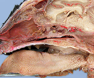
tongue (root, body, apex) [palpate]
papillae: (filiform, conical, fungiform, foliate & vallate)
lingual frenulum [palpate]
lyssa (difficult to find in the cat)
sublingual caruncle [palpate]
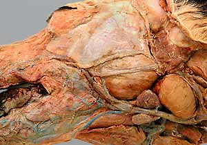
mandibular salivary duct
major sublingual salivary duct
mandibular salivary gland (named submandibular in human) [palpate]
sublingual salivary gland (monostomatic gland)
parotid salivary gland
parotid duct [palpate opening]
buccal salivary gland (prominant only in the cat)
palate [palpate]
incisive papilla [palpate] & incisive ducts
vomeronasal organ (difficult to find; need not identify)
Pharynx:
oropharynx [palpate]
palatoglossal arch [palpate]
palatine tonsil [palpate]
semilunar fold
nasopharynx
palatopharyngeal arch
auditory tube
laryngopharynx
pharyngoesophageal limen (border)
Pharyngeal mm. (identify approximate muscle locations)
cricopharyngeus m.
thyropharyngeus m.
hyopharyngeus m.
Note:
palpebra [Latin = eyelid] = eyelid
buccal [from Latin: bucca = cheek] = pertaining to cheek
buccinator [Latin = trumpeter] = muscle of the cheek
scutiform [Latin: scutum = shield & forma = form] flat cartilage in the rostral muscles of the ear
Instructor Commentary:
In quadrupeds such as domestic mammals, directional terms are different in the head compared to the rest of the body. The term "cranial" is replaced with "rostral" (Latin: rostrum = beak) because the cranium is situated in the caudal part of the head.
While you may continue to use "dorsal/ventral", you can also use "superior/inferior" in the head because, in the head, the terms mean the same in humans and quadrupeds. (In the human trunk, "superior/inferior" equates to "cranial/caudal" in quadrupeds.)
You can also use "anterior/posterior" in the head because the terms are consistent in humans and quadrupeds. (In the human trunk, "anterior/posterior" equates to "ventral/dorsal" in quadrupeds.)
Human anatomists use "superior/inferior" and "anterior/posterior" throughout the body. In veterinary anatomy we refrain from using these terms because their meanings are different, except in the head where human and veterinary meanings are the same.
The term "mouth" [Latin: os & oris] is commonly used to refer to the opening of the oral cavity (as in "close your mouth"). Anatomically, however, "mouth" refers to the oral cavity, which contains the tongue and teeth.
Dissection Steps:
Click to view a PDF list of dissection procedures for this lab:
Show List of Dissection Steps (PDF)
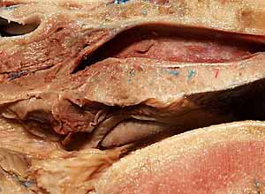
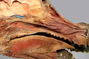
Dissection Images:
Note: Click an image to see it enlarged, view its caption, and toggle its labels.
| 1 | 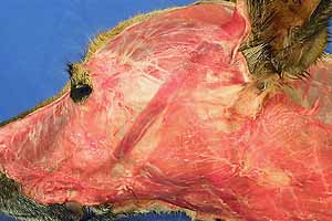 |
 |
2 |
| 3 | 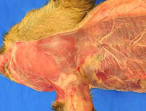 |
 |
4 |
| 5 | 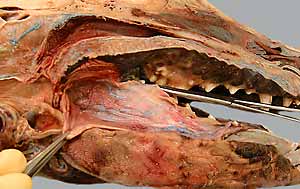 |
 |
6 |
| 7 | 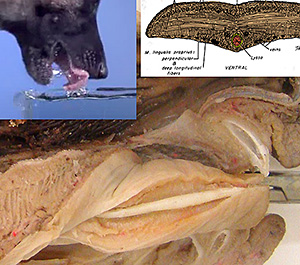 |
 |
8 |
| 9 | 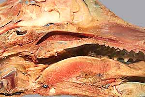 |
 |
10 |
| 11 | 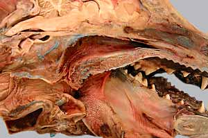 |
 |
12 |