
LAB 24 Introduction
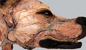
Eye and Related Structures
Superficial Veins
(Guide to the Dissection of the Dog, 8th ed., pp. 254-260)
CONTENTS:
Lab Objectives:
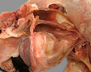
• Examine the orbit and its contents:
- eyeball and optic nerve
- muscles, vessels, and sensory & motor nerves
- lacrimal gland, fat, and the deep part of the third eyelid
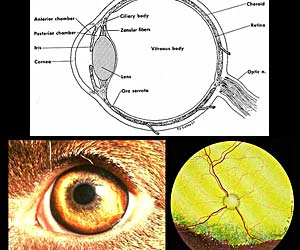
• Identify features of the eyeball:
- fibrous layer (cornea & sclera)
- vascular layer (iris, ciliary body, & choroid)
- retina and optic disc
also, aqueous humor chambers, lens, vitreous body
• Superficial veins of the head:
- external jugular v. (from maxillary v. & linguofacial v.)
- linguofacial v. (from lingual v. & facial v. )
also, observe angularis oculi v.
Anatomical Terms:
Eye & adnexia
orbit [palpate]
periorbita
lacrimal gland
superficial gland of the third eyelid
Muscles:
levator palpebrae superioris m.
rectus muscles (dorsal, ventral, medial, & lateral)
retractor bulbi m.
ventral oblique m.
dorsal oblique m.
trochlea
Eyeball:
bulbus oculi (eyeball) [palpate]
external fibrous coat
cornea [palpate]
sclera [palpate]
limbus (corneoscleral junction)
middle vascular coat (uvea)
iris [palpate]
pupil [palpate]
choroid
tapetum lucidum
ciliary body
ciliary processes
zonule (zonular fibers)
internal coat (retina)
ora serrata (margin of the optic part of the retina)
fundus
optic disk
lens
anterior [palpate] & posterior chambers
aqueous humor
vitreous chamber
vitreous body
Head: superficial veins
external jugular vein
linguofacial vein
lingual vein
facial vein
dorsal nasal v.
angularis oculi v.
maxillary vein
Note:
uvea [from Latin: uva = grape] = the vascular layer of the eyeball
uvula [Latin = little grape] = small mass on the human soft palate
vitreous [Latin: vitreus = glassy] e.g.. vitreous body
hyaline [Greek:hyalos = glass] e.g., hyaline cartilage
humor [Latin = a liquid] e.g., aqueous humor
Instructor Commentary:
Seven muscles attach to the eyeball of domestic mammals (four recti, two obliques, and one retractor). The purpose of the retractor bulbi m. is to force the third eyelid across the surface of the cornea as a protective mechanism. Humans lack a third eyelid and a retractor bulbi m., and have only six muscles attaching to each eyeball.
The gland associated with the third eyelid is called "superficial" even though it is located deep because some species, such as the pig, have two glands, and the second is still deeper than the superficial gland of the third eyelid.
Dissection Steps:
Click to view a PDF list of dissection procedures for this lab:
Show List of Dissection Steps (PDF)
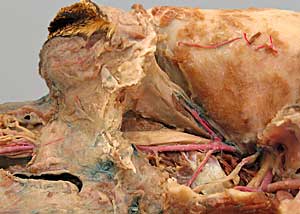
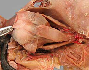
Dissection Images:
Note: Click an image to see it enlarged, view its caption, and toggle its labels.
| 1 | 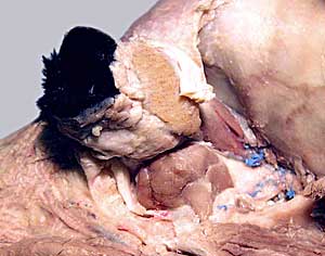 |
 |
2 |
| 3 | 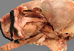 |
 |
4 |
| 5 | 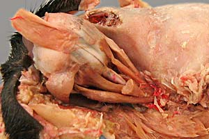 |
 |
6 |
| 7 | 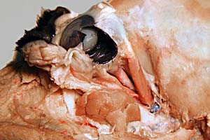 |
 |
8 |
| 9 | 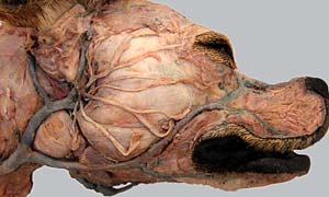 |
 |
10 |