Vestibular Nerve Action Potentials
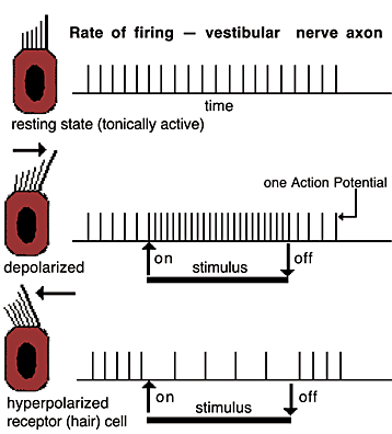
Vestibular nerves continually convey action potential (AP) traffic. In the absense of head acceleration, AP frequency is balanced in right and left vestibular nerves. Head acceleration in one direction increase AP frequency on that side and decreases frequency on the other side. Visualize this by moving the tilt knob in the following animation, developed by Kurtis Scaletta, Digital Media Center, University of Minnesota.
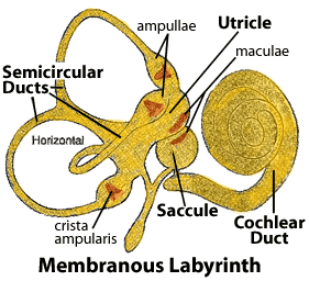
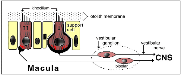
 Vestibular nerve axons innervate the vestibular apparatus. The axons synapse on receptor cells (hair cells) located within the crista ampullaris of a semicircular duct or located within the macula of the saccule or utricle of the membranous labyrinth. Head acceleration results in displacement of cilia, which leads to hair cell depolarization, release of neurotransmitter, and generation of APs in vestibular nerve axons.


Cilia displacement toward the kinocilium increases hair cell depolarization and thus AP frequency in vestibular nerve axons. Cilia displacement away from the kinocilium decreases depolarization and AP frequency. Since the vestibular apparatus of the right side is a mirror image of the left one, head acceleration in any direction displaces cilia toward kinocilia on one side and away from kinocilia on the other side.
The imbalance of AP frequency in vestibular nerves triggers reflexes that counteract head acceleration and normally keep the eyes, head, and body properly oriented. When the AP frequency imbalance is due not to acceleration but due to a unilateral lesion, then the resultant vestibular reflexes produce a vestibular syndrome: head tilt and falling toward the damaged side and eye nystagmus with the slow phase directed toward the lesion side.
© 2005
T.F. Fletcher (fletc003@umn.edu)
(Free for Educational Use)
Visit: Minnesota Veterinary Anatomy Web Site
Supported by University of Minnesota College of Veterinary Medicine
Go Top