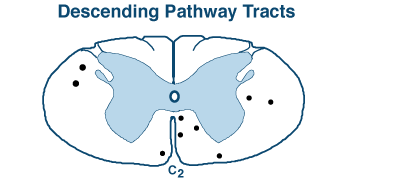
Clinical Neuroanatomy

Lower Motor Neuron

A lower motor neuron is, simultaneously, a:

    1. somatic efferent neuron, located in a cranial nerve motor nucleus or in a motor nucleus within the spinal cord ventral horn

    2. motor unit neuron that innervates a group of muscle fibers/cells within a skeletal muscle; a muscle contracts in multiples of motor units.

    3. final common pathway neuron, responsible for muscle contraction, driven by reflex activity and/or by voluntary actions

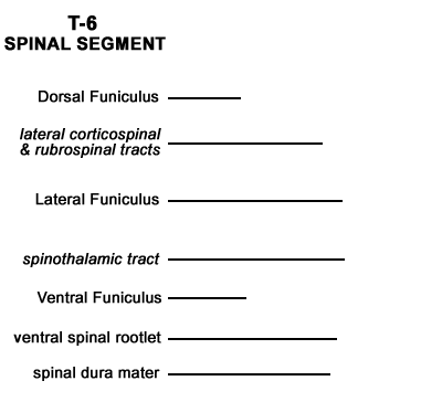
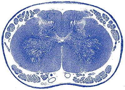
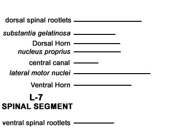
   • Spinal lower motor neurons give rise to axons that run in spinal ventral roots & spinal nerves

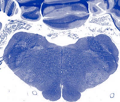
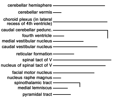
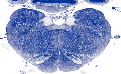
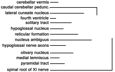
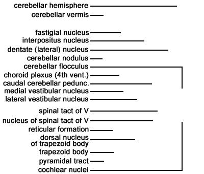
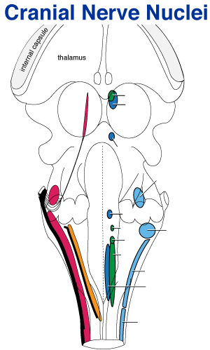
   • Cranial nerve lower motor neurons send axons into cranial nerve roots attached to the brainstem:

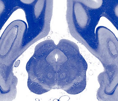
      - midbrain -- oculomotor & trochlear nerves.

      - pons -- trigeminal nerve.

      - medulla oblongata – abducent (VI) through hypoglossal (XII) nerves.

Lower motor neuron destruction: results in flaccid paralysis (loss of both voluntary and reflex movements), plus severe muscle atrophy.

Neuroanatomy Menu

Clinical Neuroanatomy

Upper Motor Neuron

Upper motor neurons are projection neurons that have cell bodies in the brain; they give rise to descending tracts that terminate on interneurons or motor neurons in cranial nerve motor nuclei or in spinal cord gray matter.

- for standing posture, descending tracts are: pontine reticulospinal & lateral vestibulospinal (both tracts originate in the hindbrain and run in the ventral funiculus of the spinal cord)

- for voluntary movements: corticospinal, rubrospinal, and medullary reticuospinal tracts are used (the first two decussate in the brain; all three descend in the dorsal half of the lateral funiculus).

Typically, voluntary movements simultaneously employ the following three tracts:

      • medullary reticulospinal tract controls axial & proximal appendicular muscles

      • rubrospinal tract, which controls proximal & middle appendicular joints

      • lateral/ventral corticospinal tracts, which control carpus/tarsus & digits (corticospinal tracts most important for raccoons, least important for ungulates)

Upper motor neuron destruction (projection neuron destruction): causes paralysis/paresis (pending the severity of damage) and often extensor hypertonus (when tracts in the ventral funiculus are spared).

(In contrast to lower motor neuron destruction, local reflexes are preserved with upper neuron destruction.)

Neuroanatomy Menu

Clinical Neuroanatomy

Spinal Cord

Spinal white matter is divided bilaterally into three regions:

   dorsal funiculus
      - contains proprioception & discriminative touch pathways (damage results in ataxia and impaired proprioceptive positioning)

   lateral funiculus
      - dorsal half: contains descussated descending tracts for voluntary movement (rubrospinal, lateral corticospinal; also, medullary reticulospinal)
      - ventral half: contains the decussated lateral spinothalamic pain pathway
         (Note: pain ascends bilaterally via multiple tracts)

   ventral funiculus
      - contains ipsilateral pathways for standing posture & extensor hypertonus (pontine reticulospinal and lateral vestibuospinal tracts)

White matter total destruction: produces voluntary paralysis, ataxia and anesthesia (caudal to the level of the lesion).

Spinal gray matter is divided bilaterally into three regions:

   dorsal horn
      - interneurons & ascending tract projection neurons that receive primary afferent input

   intermediate substance (located lateral to the central canal)
      - contains preganglionic visceral efferent cell bodies: in intermediolateral nucleus & lateral horn ( T1 to L4) and sacral prasympathetic nucleus (S1-3)

   ventral horn
      - somatic efferent neuron cell bodies within medial & lateral motor nuclei (lateral nuclei are found in enlargement segments and supply limb muscles)

Gray matter destruction: produces lower motor neuron signs (flaccid paralysis & muscle atrophy).

Neuroanatomy Menu

Clinical Neuroanatomy

Hindbrain

Brainstem: pons & medulla oblongata --


- generates standing posture via the pontine reticulospinal tract & lateral vestibulospinal tract (both tracts descend in the spinal ventral funiculus); also, corticospinal and rubrospinal descending tracts for voluntary movement run through the hindbrain

- vestibular reflexes: vestibular nuclei reflexly maintain balanced posture of eyes, head, & limbs

- cranial nerve nuclei and roots: trigeminal through hypoglossal cranial nerves attach to the hindbrain

- visceral reflex centers (cardiovascular, respiratory, gastrointestinal, urinary, etc)


Cerebellum --

- vestibular role:
      the flocculonodular lobe interacts with brainstem vestibular nuclei

- muscle tone:
      cortical damage leading to an uninhibitied fastigial nucleus produces opisthotonus

      fastigial nucleus damage results in hypotonus (weakness)

- movement synergy:
      cerebellar hemisphere damage produces disturbance of movement amplitude & strength: hypermetria, ataxia, trunk swaying, intention tremor (especially of the head)

- distance-rate-of-change perception:
      damage impairs visual menace response & avoidance of running into walls, etc.

Neuroanatomy Menu

Clinical Neuroanatomy

Midbrain

The midbrain contains:

- reticular activating system, which provides background excitation to the cerebral cortex to preclude coma

- neuromodulation nuclei within the midbrain (& pons) influence mood, movement, pain sensitivity; they widely distribute dopamine, serotonin, acetylcholine & norepinephrine.

- red nucleus, gives rise to axons that immediately decussate and descend as the rubrospinal tract. In quadrupeds, it is the major tract for voluntary movement (including suppression of standing posture during limb flexion). Pending its location, a unilateral lesion may cause ipsilateral (rubrospinal tract) or contralateral (red nucleus) paresis/paralysis

- corticospinal fibers run through the midbrain, prior to decussating at the brain-spinal cord juncture

- cranial nerve nuclei and roots:

      oculomotor nerve (enables eye movement medially & pupil constriction)

      trochlear nerve (innervates dorsal oblique m.)

- tectum: orientation of head, eyes & ears toward a sudden stimulus, either visual (rostral colliculus) or auditory (caudal colliculus)

- pretectal region: pupillary light reflex

- periaqueductal gray: relays visceral information and gives rise to an endogenous analgesia system that suppresses pain pathway traffic

Neuroanatomy Menu

Clinical Neuroanatomy

Diencephalon

The diencephalon includes the thalamus, hypothalamus, subthalamus, and epithalamus.

- thalamic nuclei: all ascending tracts destined for the neocortex relay in a thalamic nucleus, including pathways from the cerebellum and axons arising from basal nuclei

- hypothalamus: autonomic control, including visceral responses related to emotional states, pituitary gland control, and visceral related responses/behavior, including: eating, drinking, temperature regulation and circadian rhythms linked to daylight

- subthalamus: used by the cerebral cortex to suppress unwanted movements (the subthalamus excites inhibitory neurons located within basal nuclei).

- epithalamus: involved in threat induced inhibition of movement (movement freeze behavior)


vision:

      optic nerve - destruction produces complete blindness of the ipsilateral eye

      optic tract - destruction produces contralateral blindness in the visual fields of both eyes

      lateral geniculate nucleus - relays conscious vision to the neocortex

      pretectal region - processes reflex pupil constriction in response to light (via oculomotor nerve)

hearing:

      medial geniculate nucleus - relays conscious hearing to the neocortex

      auditory pathways are bilateral in the brain beyond cochlear nuclei

Neuroanatomy Menu

Clinical Neuroanatomy

Cerebrum

The cerebrum is composed of two cerebral hemispheres, each containing white and gray matter. Cerebral gray matter is either superficial (cerebral cortex) or buried (basal nuclei). The dorsal cerebrum is covered by six-layered neocortex.

The term rhinencephalon refers to the older, ventral cerebrum that is concerned with olfaction, emotions, and processing recent memory.


- neocortex is essential for consciousness & mental alertness, intelligence & learning, pattern recognition & cognition, self-awareness & comprehension of significance, memory storage & recall, planning & appropriate behavior, and selecting & executing voluntary movement.

- neocortical regions:

primary sensory areas (somatosensory, visual, auditory, etc.) - receive thalamic sensory projections

sensory association cortex (surrounds primary areas) - extracts pattern/significance from sensations

prefrontal association cortex (frontal pole) - plans/directs goal-oriented focused behavior

premotor cortex - involved in movement selection and motor learning

motor cortex - somatotopic organization of joint movements; drives pyramidal/extrapyramidal tracts.

- some basal nuclei (caudate, putamen, & globus pallidus) participate in inhibitory circuits that suppress undesired movement and enable desired voluntary movement.

- limbic system structures (e.g., cingulate gyrus, septum, amygdala, hippocampus) process emotional reactions that drive memory and behavior (e.g., self-defense, escape, mating, etc.). Limbic system also includes components of the diencephalon and midbrain.

- olfactory neurons synapse in the olfactory bulb, which sends axons to the piriform lobe for conscious olfaction and to the limbic system for emotional responses (e.g., pheromones elicit emotionally driven behavior).

Neuroanatomy Menu