|
|
|
 |
|
Neuroglia - Image 1:
Three types of CNS gliocytes are drawn as they would appear with a Golgi stain. Astrocytes are relatively large. Oligodendrocytes form myelin sheaths in the CNS. Microglia are relatively rare in normal CNS tissue.
|
|
|
|
 |
|
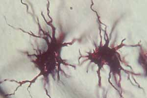
Neuroglia - Image 2:
Two fibrous astrocytes are shown (Golgi stain).
|
|
|
|
 |
|
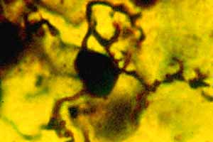
Neuroglia - Image 3:
Oligodendrocytes are shown in CNS white matter (Golgi stain). |
|
|
|
 |
|
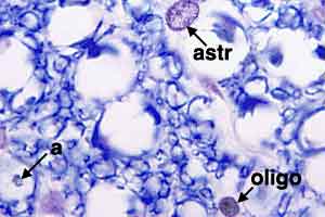
Neuroglia - Image 4:
Spinal cord white matter, stained with Luxol Blue. Typical astrocyte nuclei (astr) are relatively large, oval, and chromatically stippled. Typical oligodendrocyte nuclei (oligo) are small, round, and packychromatic. Myelinated axons (a) are evident within vacuoles produced by lipid extraction during tissue processing. |
|
|
|
 |
|
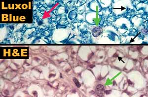
Neuroglia - Image 5:
Spinal cord white matter, stained with Luxol Blue (top) and H&E (bottom). Typical nuclei of astrocytes (green arrows) and oligodendrocytes (red arrows) can be seen. Myelinated axons are also evident (black arrows). |
|
|
|
 |
|
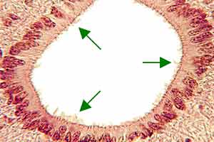
Neuroglia - Image 6:
Spinal cord central canal (H&E stain). The central canal of the spinal cord is lined by ependymal cells which have cilia (arrows) that project into the lumen of the central canal. |
|
|
|
 |
|
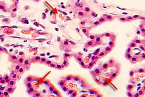
Neuroglia - Image 7:
Choroid plexus of a brain ventricle (H&E stain). Each choroid plexus consists of vascular villi lined by cuboidal ependymal cells, called choroid plexus epithelium. The arrows indicate red blood cells in vessels. |
|
|
|
 |
|
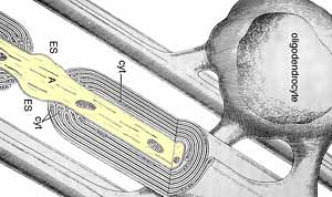
Neuroglia - Image 8:
An ultrastructural drawing of a CNS oligodendrocyte contributing myelin internodes to several axons. A myelin sheath is interrupted by nodes. Myelin internodes are formed by wrappings of oligodendrocyte plasma membrane.
|
|
|
|
 |
|
Neuroglia - Image 9:
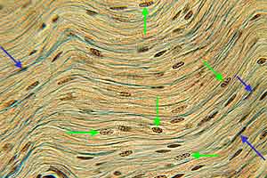
Bovine sympathetic trunk, longitudinal section (Triple stain). Numerous lemmocyte nuclei (green arrows) are visible in this PNS nerve. Some of the nuclei belong to endoneurial fibrocytes (blue arrows).
|
|
|
|
 |
|
Neuroglia - Image 10:
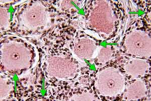
Canine spinal ganglion (H&E stain). Unipolar neuron cell bodies are surrounded by satellite glial cells (arrows).
|
|
|
|
 |
|
Neuroglia - Image 11:
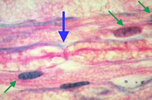
Canine peripheral nerve, longitudinal section (Triple stain). The green arrows indicate lemmocyte nuclei. The blue arrow indicates a myelin node. The pink, foamy material is myelin neurokeratin. |
|
|
|
 |
|
Neuroglia - Image 12:
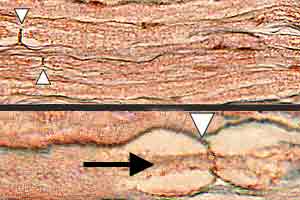
Canine peripheral nerve, longitudinal section (Triple stain). Top: several myelin nodes (white arrows) are visible at low power. Bottom: A myelin node is shown at high power. Black arrows indicate axons. Neurokeratin (nk) is labeled. |
|
|
|
 |
|
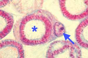
Neuroglia - Image 13:
Canine peripheral nerve, transverse section (Triple stain). An axon (asterisk) is surrounded by a myelin sheath, in turn surrounded by lemmocyte cytoplasm and the lemmocytes nucleus (arrow)
|
|
|
|
 |
|
Neuroglia - Image 14:
An ultrastructural drawing of a transverse section through a myelinated nerve fiber. The axon (A) is surrounded by myelin and then by lemmocyte cytoplasm and lemmocyte nucleus (N), all within a basal lamina. Ans inset shows myelin at high magnification. |
|
|
|
 |
|
Neuroglia - Image 15:
A drawing of the ultrastructure of a myelin node from the CNS (left) and the PNS (right). The term paranode refers to the cytoplasmic region of the myelin sinternode that is adjacent to the node. |
|
|
|
 |
|
Neuroglia - Image 16:
An ultrastructural drawing of a transverse section through a lemmocyte ensheathing many non-myelinated axons. Each axon (A) is encased in a protected space formed by lemmocyte processes. |
|
|
|
 |
|
Neuroglia - Image 17:
A schematic illustration of the developmental relationship of neurolemmocytes to myelinated and non-myelinated axons. Initially, each lemmocyte embraces multiple axons. For non-myelinated axons, a lemmocyte ensheathes many axons individually (lower left). For myelinated axons, a lemmocyte rotate processes around each axon to produce a myelin sheath internode.
|