|
|
|
 |
|
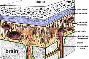
Meninges - Image 1:
Artist illustration of cranial meninges. Dura mater, the most superficial menix, also functions as periosteum for bones of the cranial cavity. Arachnoid is applied to the deep surface of dura mater. Arachnoid trabeculae connect to pia mater. Pia mater, the deepest menix, is attached to a glial limiting membrane formed by surface astrocytes of the brain. |
|
|
|
 |
|
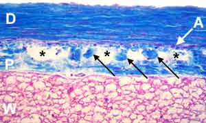
Meninges - Image 2:
Histologic section through spinal meninges (Triple stain). Dura mater (D) is dense collagenous tissue. Arachnoid (A) consists of flattened fibrocytes attached to the deep surface of dura mater. Pia mater is composed of collagen fibers along the surface of white matter (W) and flattened fibrocytes lining the subarachnoid space (asterisk). |
|
|
|
 |
|
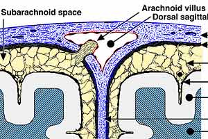
Meninges - Image 3:
Schematic illustration of cranial meninges including an arachnoid villus. Meninges are labeled, including the subarchnoid space which is filled with cerebrospinal fluid and traversed by arachnoid trabeculae. Along the midline, where the falx cerebri partition arises, a dorsal sagittal venous sinus is located. |
|
|
|
 |
|
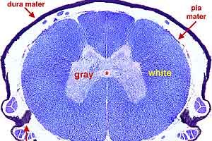
Meninges - Image 4:
Transverse section through a spinal cord segment and meninges (Luxol Blue stain). Pia mater is attached along the white matter surface. Dura mater (D) is the most superficial menix. (An epidural space separates dura mater from periosteum lining the vertebral canal.) Arachnoid lines the dura mater. Bilaterally, a denticulate ligament extends from pia mater to dura mater. |
|
|
|
 |
|
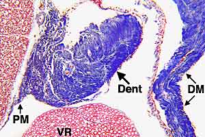
Meninges - Image 5:
High power view of the denticulate ligament (Dent) (Triple stain). Pia mater (PM) is attached to the surface of spinal white matter. The denticulate ligament is an expansion of pia mater collagen that periodically sends extensions toward the dura mater (DM). |