|
|
|
 |
|
Neuron - Image 1:
A sketch illustrating functional regions of a multipolar neuron. Differences in plasma membrane protein constitutes the basis for functional specialization within an individual neuron. |
|
|
|
 |
|
Neuron - Image 2:
A drawing of the three categories of neurons, based on the number of processes emanating from the cell body: unipolar, bipolar, and multipolar. Multipolar neurons come in a variety of shapes. |
|
|
|
 |
|
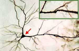
Neuron - Image 3:
A pyramidal neuron from a canine cerebral cortex (Golgi stain). The multipolar cell body (arrow) is shaped like a pyramid, with apical and basal dendrites. The inset shows part of the apical dendrite, enlarged so that dendritic spines can be seen. |
|
|
|
 |
|
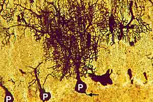
Neuron - Image 4:
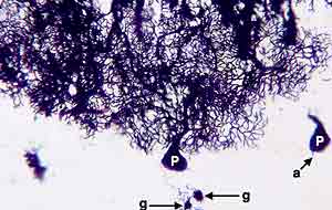
Purkinje neuron from a canine cerebellar cortex (Golgi stain). Three Purkinje (piriform) cells are more or less stained. The cell body of the Purkinje neuron is pear-shaped (P). A dense dendritic tree can be seen entering the molecular layer of the cerebellar cortex. |
|
|
|
 |
|
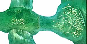
Neuron - Image 5:
Neurons of the cerebellar cortex are shown (Golgi stain). A massive dendritic tree emanates from the cell body (P) of a Purkinje cell (neuron). Two granule cells (g) are stained. |
|
|
|
 |
|
Neuron - Image 6:
Three multipolar neuron cell bodies from ventral horn gray matter of the spinal cord are shown (Nissl stain). Typical features of neuronal perikaria include a prominent nucleolus within a large leptochromatic nucleus and clumps of Nissl substance (rER & polyribosomes). |
|
|
|
 |
|
Neuron - Image 7:
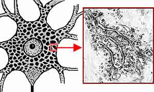
A drawing of a multipolar neuron illustrating cytoplasmic Nissl bodies. The insert shows the ultrastructure of Nissl substance; <i>viz.,</i> clumps of rough endoplasmic reticulum (rER) and polyribosomes. |
|
|
|
 |
|
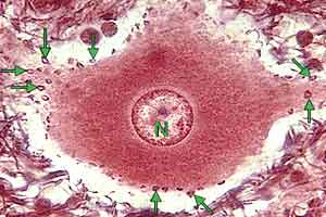
Neuron - Image 8:
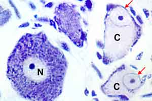
Two chromatolytic cell bodies (C) are adjacent to a normal cell body (N). Features of neuron chromatolysis include: a swollen cell body, eccentric nucleus, and loss of Nissl substance except along the margins of the cell body. |
|
|
|
 |
|
Neuron - Image 9:
A drawing illustrating ultrastructural features of a neuron cell body is shown. Four types of synapses are included. |
|
|
|
 |
|
Neuron - Image 10:
Ventral horn motor neuron (thick section, stained with a silver). Synapses are evident as dark circles (arrows) along the margin of the cell body. |
|
|
|
 |
|
Neuron - Image 11:
Schematic illustration of the ultrastructure of a sectioned axo-dendritic synapse. A yellow pre-synaptic terminal bulb (bouton) is separated by a synaptic cleft from a dendrite (brown). |
|
|
|
 |
|
Neuron - Image 12:
An artist drawing of two axo-dendritic synapses is presented. The left illustrates a synpase in passing (en passant). The right involves a terminal bulb. Synaptic vesicles are shown in white. |
|
|
|
 |
|
Neuron - Image 13:
Artist drawing of three neuromuscular synapses. The pre-synaptic element of a neuromuscular synapse consists of a circumscribed cluster of terminal branches that collectively constitute an end plate. |
|
|
|
 |
|
Neuron - Image 14:
Illustration of the ultrastructure of a neuromuscular synapse. The pre-synaptic element is filled with synaptic vesicles. The surface area of the synaptic cleft is greatly increased by junctional folds. |