CNS - Image 9
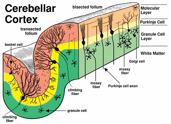
An artist drawing of the cytology and circuitry of the cerebellar cortex is presented. A cerebellar folium is shown in transverse section (left) and longitudinal section (right). The outer molecular layer (brown) has dendrites and axons but few cell bodies. The Purikinje cell layer (yellow) contains large Purkinje (piriform) neurons. Notice that Purkinje neurons have a broad dendritic tree with its width oriented perpendicular to the long axis of the folium. The granule cell layer (green) contains a dense accumulation of very small neurons. Climbing fibers and mossy fibers enter the cerebellar cortex. Axons of Purkinje neurons leave the cerebellar cortex.