Meninges - Image 3
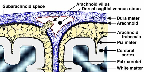
Schematic illustration of cranial meninges including an arachnoid villus. Meninges are labeled, including the subarchnoid space which is filled with cerebrospinal fluid and traversed by arachnoid trabeculae. Along the midline, where the falx cerebri partition arises, a dorsal sagittal venous sinus is located. Mostly microscopic arachnoid villi project into the venous sinus and function as one-way valves for cerebrospinal fluid drainage into the blood stream.