Spinal Ganglia
Ganglia are enlargements of periperal nerves produced by accumulations of neuron cell bodies. Unipolar cell bodies are found in spinal ganglia (on dorsal roots) and in ganglia on the roots of cranial nerves. There are no synapses within these sensory ganglia. Multipolar neurons (postganglionic autonomic neurons) are found in autonomic ganglia (slide 56, Histology Box). Preganglionic autonomic neurons synapse on the postganglionic neurons within autonomic ganglia.
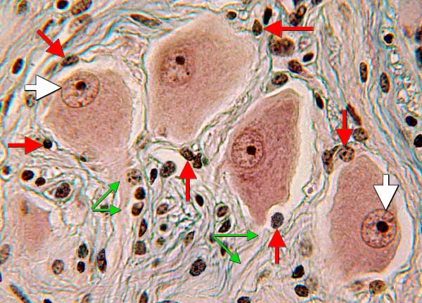
On glass slide 55 in your Histology slide box, spinal ganglion of a cow (H&E stain), find unipolar cell bodies and satellite lemmocytes as illustrated below.

Autonomic Ganglia:
Autonomic ganglia are found within autonomic nerves. The ganglia contain postganglionic visceral efferent neurons that receive synaptic input from preganglionic visceral efferent neurons. As shown below (triple stain), an autonomic ganglion contains multipolar neuron cell bodies with eccentric nuclei (white arrows); axons (green arrows) arise from each cell body. Satellite glial cells [also called amphicytes] (red arrows) are sparse and form poorly-defined capsules around individual cell bodies:
