Neuroglia
Neuroglial cells are the supportive cells of nervous tissue. They outnumber neurons about 10 to 1. Like neurons, glial cells are composed of cell bodies and cell processes. (Note: Glial processes are visible only in special stained preparations, such as a Golgi stain.)
Three major types of neuroglial cells are recognized in the central nervous system:
�
(a) astrocytes -- provide structural & functional support for the CNS;
�
(b) oligodendroglia -- form myelin in the CNS, and
�
(c) microglia -- serve as a macrophage in the CNS.
�
Astrocytes:
On glass slide 49 in your Histology slide box, cerebral cortex of dog (Golgi stain), search for astrocytes such as those illustrated below.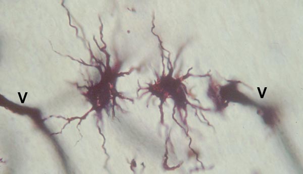 �
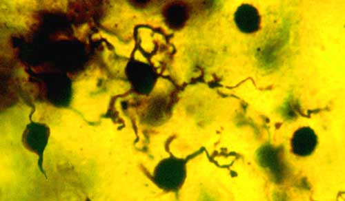
�Oligodendrocytes:
On glass slide 49 in your Histology slide box, cerebral cortex of dog (Golgi stain), search for ologodendrocytes such as those illustrated below.
 �
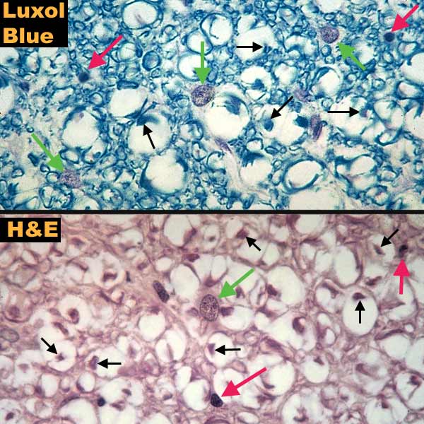
�On glass slide A-1 in your Neuroanatomy slide box, canine spinal cord, search the white matter for astrocyte and ologodendrocyte cell bodies as they appear in routine stains, illustrated below. �
 �
��
Microglia:
Microglia are difficult to find. Microglial cells have small elongate perikarya and short cell processes. They comprise only about 4% of the glial cell population under normal circumstances. An example of a microglial cell will be on demonstration in the laboratory.
�