Peripheral Nerves
Peripheral nerves consist of bundles of myelinated and non-myelinated nerve fibers enveloped by connective tissue.
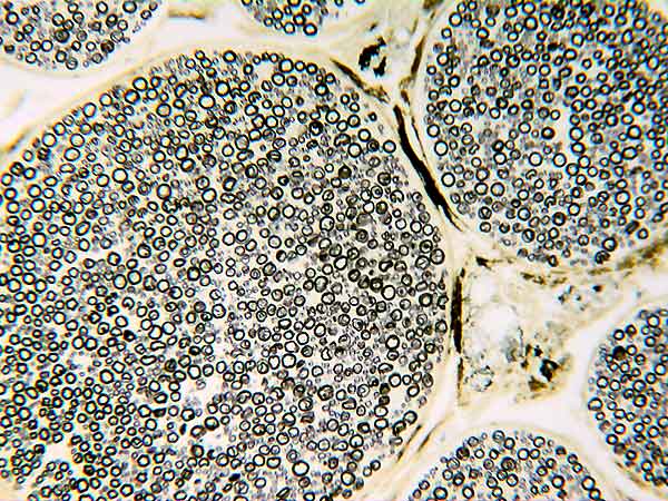
On glass slide 51 in your Histology slide box, longitudinal and transverse sections of a sciatic nerve from a horse are evident (triple stain). In the transverse section, find epineurium, perineurium, and endoneurium (see Figs. 7 & 8).
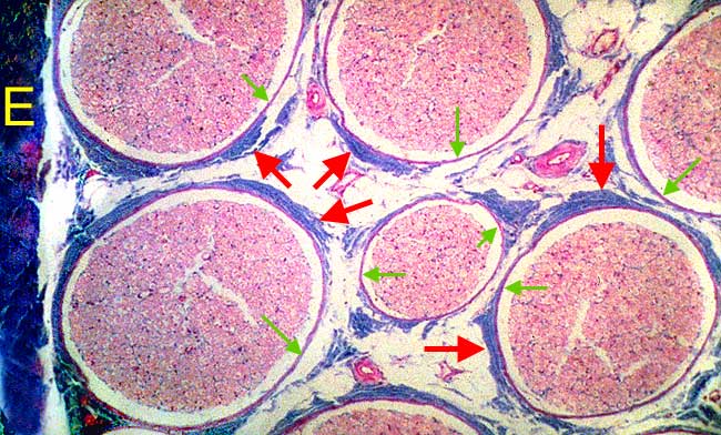 �
�Fig. 7. In this view of a transected equine sciatic nerve, epineurium (E) is present on the left side of the field. Fibrous perineurium (red arrows) surrounds individual nerve fascicles. Notice perineural epithelium (green arrows) deep to the fibrous perineurium (see Fig. 8). The endoneurium surrounding individual myelinated fibers within the fascicle is barely evident at this magnification.
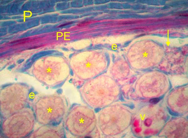 �
�Fig. 8. In this enlarged view of a nerve fascicle shown in Fig. 7, identify fibrous perineurium (P) at the top. Deep to the fibrous perineurium, perineural epithelium (PE) is composed of several layers of flattened cells derived from mesoderm. Perineural epithelial cells (perineural epithelioid cells) form a protective barrier around the nerve fascicle. Within the fascicle, sparse endoneurium (e) is evident surrounding individual myelinated fibers. Axons (asterisks) of myelinated fibers are visible, as is a blood vessel (V).
Profiles of non-myelinated axons (ensheathed by lemmocytes) are evident as clustered dots (arrow).
Profiles of non-myelinated axons (ensheathed by lemmocytes) are evident as clustered dots (arrow).
Myelinated nerve fibers:
On glass slide 51 (triple stain), search the transverse section of the equine sciatic nerve under medium power for evidence of lemmocyte cytoplasm surrounding a myelin sheath, see below.
 �
�Fig. 9. Myelinated fibers are shown in cross section. In the center, a myelinated axon (asterisk) is surrounded by a myelin sheath which, in turn, is surrounded by lemmocyte cytoplasm. Myelin appears as neurokeratin, the myelin protein residue that remains following lipid extraction during tissue processing.
Also on glass slide 51 (longitudinal and transverse sections of an equine sciatic nerve), search the longitudinal section at high power for evidence of a myelin node (illustrated below).
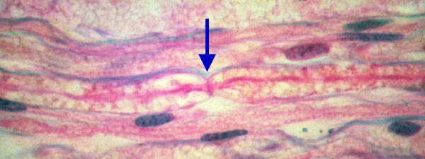 �
�Fig. 10. In this longitudinal section, a myelin node (of Ranvier) is evident at the center (arrow). Notice the axon (dark band) continuing through the juncture of adjacent myelin sheaths where myelin neurokeratin surrounds the axon. (Neurokeratin represents a myelin sheath artifact resulting from lipid extraction during tissue processing.) Nuclei in the section belong to lemmocytes, although some belong to fibrocytes of the endoneurium.
 �
�Fig. 11. Transverse section throung a canine vagus nerve. Only myelin sheaths (plus some lipid associated wth epineurium and perineurium) are stained. Notice the rage in size of myelinated fibers. Within a fascicle, the white gaps between myelinated fibers are occupied by non-myelinated fibers.
�
Non-myelinated nerve fibers:
On glass slide 52 in your Histology slide box, sympathetic trunk from a cow, most of the nerve fibers are non-myelinated.
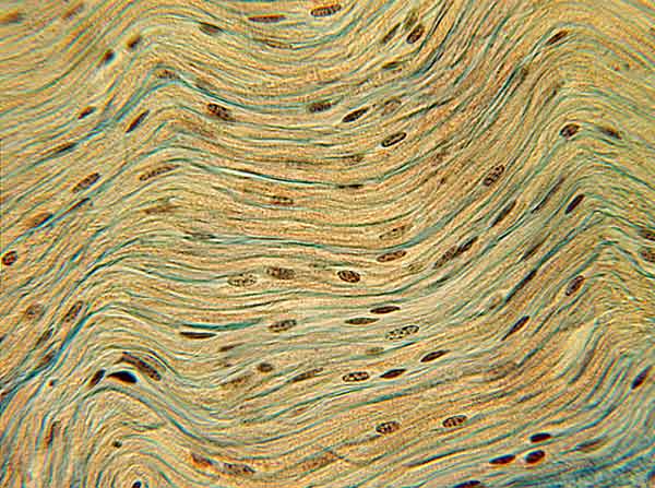 �
�Fig. 12. Although some myelinated preganglionic fibers are present, non-myelinated postganglionic fibers predominate in this longitudinal section through a bovine sympathetic trunk. Non-myelinated fibers are thin and they blend with lemmocyte cytoplasm. Notice the high density of lemmocyte nuclei, the wavy pattern of nerve fibers, and evidence of connective tissue sheaths.
�
Go Top