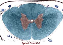
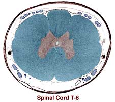
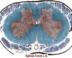
Spinal gray matter is butterfly-shaped. It extends from the ependymal cells lining the central canal to the surrounding white matter. Spinal gray matter is divided bilaterally into dorsal horn, intermediate substance, and ventral horn. At thoracolumbar levels, intermediate substance features a lateral horn. Intermediate substance lacks precise boundaries; in general, it is around the central canal and between dorsal and ventral horns.
Spinal neurons within the gray matter are either efferent neurons (axons enter ventral roots), projection neurons (axons join white matter tracts), or interneurons (axons remain within gray matter). The gray matter "horns" are actually profiles of gray columns. Columns of neuron cell bodies, when transected, appear as clusters of neuron cell body profiles within gray matter. The cell body clusters are called nuclei. Some nuclei (columns of cell bodies) are present throughout the spinal cord, other nuclei have more restricted segmental distributions.
 The dorsal horn surface is capped by a marginal nucleus (lamina I) which is thin and not distinct in transverse sections. A population of small neurons forms a very distinctive substantia gelatinosa (lamina II). The remainder of the dorsal horn may be considered nucleus proprius. The nucleus thoracicus, located medially in the base of the dorsal horn, is present in thoracolumbar segments; axons from the nucleus form the dorsal spinocerebellar tract (Note: nucleus thoracicus projection neurons are large but sparse and not evident in some sections).
 In the intermediate substance, one nucleus is found only in thoracic and cranial lumbar segments of the spinal cord. The intermediolateral nucleus, which forms a lateral horn, is composed of sympathetic preganglionic neurons.
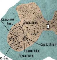
The ventral horn contains somatic efferent motor neurons. Medial motor nuclei innervate muscles of the trunk and are found in all spinal segments. Lateral collections of motor neurons, which innervate limb muscle, are seen in segments of the cervical and lumbosacral enlargements. Motor nuclei are somatotopically arranged.
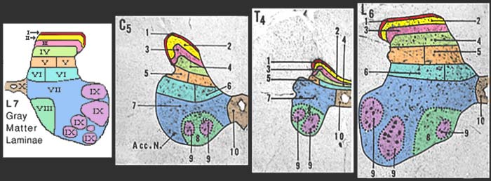
Much of the spinal gray matter is outside of recognizable nuclei. An alternative of method of categorizing gray matter involves defining spinal laminae (below), as opposed to nuclei. Laminae offer the advantage of including all regions of gray matter. The disadvantage of laminae is that, unlike nuclei, laminar boundaries are not evident in normal sections.
Below are links for viewing spinal gray matter in better quality sections.