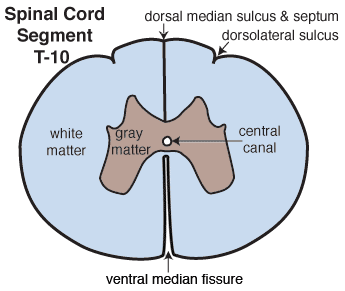
 A drawing of a transverse section through the T10 spinal segment is shown on the right. The spinal cord is bilaterally symmetrical. Ventrally, the halves are separated by a ventral median fissure (into which pia mater invaginates). Dorsally, spinal halves are demarcated superficially by a dorsal median sulcus. Deep to the sulcus, a dorsal median septum (caudally a fissure) separates the halves. Bilaterally a dorsolateral sulcus marks the entry site of dorsal rootlets into the spinal cord. At the center of the section, the central canal is lined by ependymal cells and surrounded by butterfly-shaped gray matter, which is surrounded by white matter.
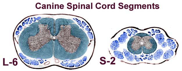
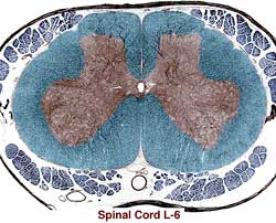
The above images (taken from glass slide A-2) are transverse sections of spinal segments L6 & S2. Notice that gray matter is butterfly-shaped surrounding the central canal, white matter is stained blue, and meninges surround the spinal cord. Roots streaming caudally to join the cauda equina are evident surrounding section S2. Click above to see all four section of slide A-2. Click below to see better quality images of spinal cord segments.
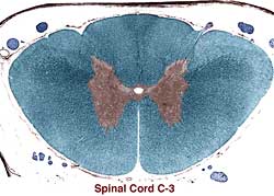
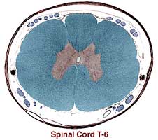
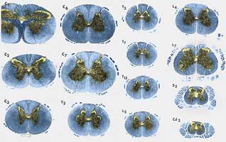 Segment Identification: On slide A-2, you could be expected to identify cervical, thoracic, lumbosacral enlargement, and sacral regions of spinal cord based on the morphologic features that you observe on the transverse sections? Click the image on the right to see spinal cord transections from multiple levels of spinal cord. Ask yourself: Why is gray matter increased in certain regions, e.g., lateral horn, lateral neurons in ventral horn? Why does white matter decrease from cranial to caudal? Notice that there are regional difference in shape of the dorsal gray horn and of the overall cord section. Why are surrounding rootlets more abundant in lumbosacral segments?