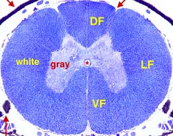
 In each half of the spinal cord, white matter is divided into three major bundles, called funiculi. The dorsolateral sulcus marks the division between dorsal funiculus and lateral funiculus. The boundary between lateral funiculus and ventral funiculus is arbitrarily set where the most lateral bundle of ventral root fibers passes transversely through the white matter. Right and left ventral funiculi are connected by a ventral white commissure, located along the midline ventral to gray matter and dorsal to the ventral median fissure.
 Spinal white matter consists of nerve fibers entering from dorsal roots; nerve fibers exiting to ventral roots; and millions of longitudinally oriented fibers organized into spinal tracts (some tracts are called fasciculi). Ascending spinal tracts convey information cranially from spinal cord projection neurons to the brain. Descending spinal tracts convey information caudally from brain neurons to the spinal cord. Although spinal tracts are anatomically nondescript (all fibers look alike); functionally, each tract is distinct since its fibers go to, or come from, a particular location in the brain and they signal a particular type of information. Some tract fibers arise and terminate entirely within the spinal cord and do not reach the brain. They are located deep, next to gray matter, and collectively they are called fasciculus proprius.
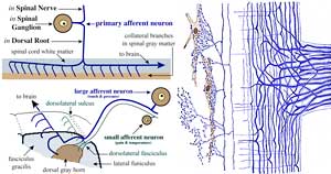 As dorsal rootlet nerve fibers enter the spinal cord, they bifurcate into cranially and caudally directed branches. These primary branches give off collateral branches that arc in a transverse plane and enter the dorsal horn gray matter. Thinly myelinated and nonmyelinated fibers (pain & temperature fibers) collect laterally as the dorsal rootlet enters the cord. Their cranial and caudal branches form the dorsolateral fasciculus. Thick myelinated fibers (touch & kinesthesia) gather medially in the dorsal funiculus as the dorsal rootlet enters the cord. Their cranial branches continue on to the brain. Such cranial branches form fasciculus gracilis (from the caudal half of the body) and fasciculus cuneatus (from the cranial half of the body).
 Within a tract, fibers are organized such that the shortest ones closest to gray matter; thus, fibers to the cervical region are deeper than those to the sacral region. In the dorsal funiculus, ascending branches of myelinated touch & kinesthesia fibers are regionally organized (Ca, S, L, T, & C). Those from the caudal half of the body constitute fasciculus gracilis. Those from the cranial half of the body form fasciculus cuneatus. The shortest tract fibers form fasciculus proprius (FP), immediately adjacent to gray matter. Lateral and ventral corticospinal tracts are depicted so as to illustrate that tracts consist of a dense core surrounded by a zone of decreasing fiber density.