Spinal Cord Anatomy
CLOSE
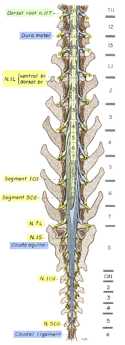
Canine Spinal Cord — Caudal Half
The caudal half of a canine vertebral column has been drawn after a laminectomy to expose the spinal cord. Spinal cord segments are labeled and locations of vertebral bodies separated by intervertebral discs are labeled to the right.
Dura mater (blue) has been removed except along the right side. Dura mater envelops spinal roots including spinal ganglia.
Notice that thoracolumbar spinal segments are long and located within nominally corresponding vertebrae. Thereafter, segments progressively shorten in length and spinal roots elongate as segments shift position cranial to nominally corresponding vertebrae. Sacral and caudal roots streaming caudally are referred to as the cauda equina. Notice that the cauda equina is initially intrathecal (within the main cylinder of spinal dura mater); thereafter, the roots are enveloped by dural sheaths in the epidural space.
The term conus medullaris refers to the cone-shaped region of spinal cord caudal to the lumbosacral enlargement (L4 — S1). The cord terminates approximately at the L6-L7 intervertebral disc. Thereafter a terminal filament of glial tissue continues for some distance. The term caudal ligament refers to the terminal filament enveloped by a dural sheath.
Go Top