Spinal Cord Anatomy
CLOSE
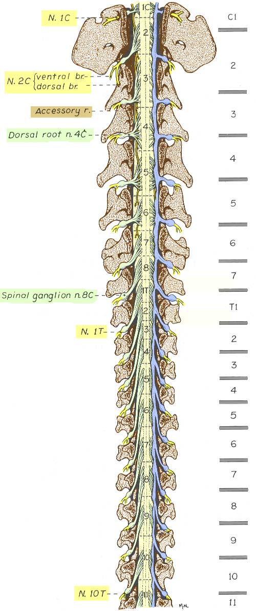
Canine Spinal Cord — Cranial Half
The cranial half of a canine vertebral column has been drawn after a laminectomy to expose the spinal cord. Spinal cord segments are labeled and locations of vertebral bodies separated by intervertebral discs are labeled to the right.
Dura mater (blue) has been removed except along the right side. Dura mater envelops spinal roots including spinal ganglia.
Notice that spinal segments vary in length and that spinal roots must elongate to reach intervertebral foramina where segments are shifted cranially. In the cervical region, notice that the spinal root of the accessory cranial nerve (Accessory r., tan) emerges laterally, between dorsal and ventral roots. The C1 spinal nerve (N.1C, yellow) exits from a lateral foramen, rather than an intervertebral foramen like other spinal nerves. The C8 spinal segment appears to be "extra" (it lacks a nominally corresponding vertebra). Thus, caudal to the cervical region, spinal nerves exit through intervertebral foramina located at caudal margins of nominally corresponding vertebrae.
The C3 segment is the longest. Thereafter, segments progressively shorten in length. After the T2 segment, segments progressively lengthen. The cervical enlargement (C6, 7, 8, & T1) which innervates the thoracic limb (brachial plexus) is centered approximately at the C6-C7 intervertebral disc.
Go Top