Spinal Cord Anatomy
CLOSE
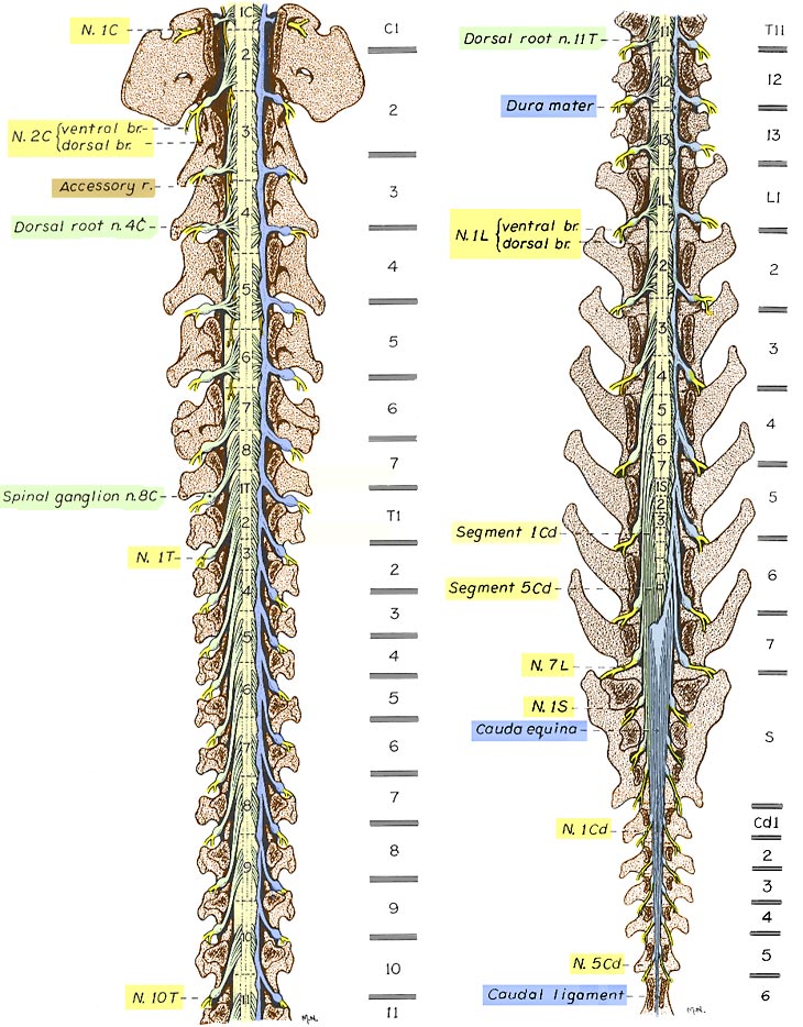
Canine Spinal Cord
Cranial and caudal halves of a canine vertebral column are illustrated, after a laminectomy to expose the spinal cord. Spinal cord segments are labeled, and locations of vertebral bodies separated by intervertebral discs are shown to the right. Dura mater (blue) has been removed except along the right side. The illustrated position relationship of spinal cord segments to vertebrae represents the most common relationship for medium and large dogs (typical variation is half a vertebral length cranial or caudal to that shown). In small dogs (under 7kg) spinal cord segments are positioned more caudally than is shown.
Go Top