The telencephalon (or cerebrum) consists of two cerebral hemispheres plus a small mid-line component called lamina terminalis. The lamina terminalis forms the rostral wall of the third ventricle and contains the rostral commissure.
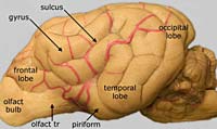
Lobes: Each cerebral hemisphere can be divided into a rostral frontal lobe, a caudal occipital lobe, a lateral, poorly developed, temporal lobe, and an ill-defined parietal lobe, located between frontal and occipital lobes.
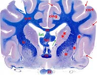
A cerebral hemisphere is composed of surface gray matter called cerebral cortex, underlying white matter and deep masses of gray matter that are collectively called basal nuclei. In addition, each hemisphere contains a cavity, the lateral ventricle. Each lateral ventricle communicates with the third ventricle though an interventricular foramen (a shared choroid plexus passes through the foramen).
Superficial gray matter of the cerebral hemisphere is called cerebral cortex (most of the cortex is neocortex, in contrast to old paleocortex of the piriform lobe). The cortical surface features sulci (grooves) and gyri (ridges). In the dog, identify the cruciate sulcus and the coronal sulcus. These sulci are landmarks for locating the primary motor area (around the cruciate sulcus) and the somatosensory area (below the coronal sulcus) of the cerebral cortex.
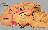
Cerebral white matter contains fibers that connect neighboring or distant gyri, fibers that communicate between hemispheres, and fibers that connect the hemisphere with the brainstem. The corpus callosum is a mass of fibers that connect right and left cerebral hemispheres. The internal capsule refers to fibers running to and from the brain stem. (To early anatomists, the fibers seemed to encapsulate the basal nuclei and the thalamus.)
|
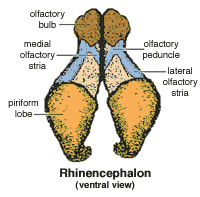
Each cerebral hemisphere can be functionally and anatomically divided into phylogenetically new vs. old components. The phylogenetically old portion is located ventrally; it is called the rhinencephalon (nose brain) because it deals with olfaction. Identify the following three components of the rhinencephalon on the sheep brain shown on the right: olfactory bulb (2), piriform lobe (7), and lateral olfactory tract (6). Click the images for more information. |

|
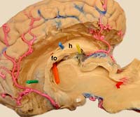
A deep component of the rhinencephalon is the hippocampus (the name reflects its resemblance to a sea horse when sectioned transversely). The hippocampus is a bilateral gray matter structure that begins deep to the piriform lobe. It curves dorsally and rostrally, like the lateral ventricle; the hippocampus forms the deep boundary of the lateral ventricle. Fibers from the hippocampus emerge laterally as a fimbria and then continue rostrally as the fornix. The fornix curves ventrocaudally to end ultimately in the midbrain.