
Lab 4 - Image 14
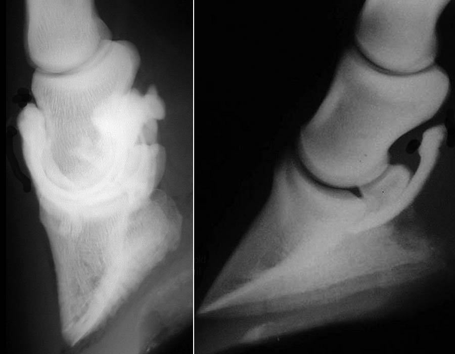
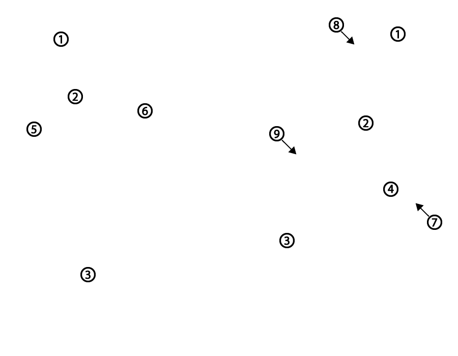
Positive contrast radiographs of the DIP joint (left) and the navicular bursa (right, courtesy E. Malone) 1, proximal phalanx = P1; 2, middle phalanx = P2; 3, distal phalanx = P3; 4, distal sesamoid (navicular) bone; 5, dorsal pouch of the distal interphalangeal (DIP) joint; 6, palmar pouch of the distal interphalangeal (DIP) joint; 7, navicular (podotrochlear) bursa; 8, proximal interphalangeal (PIP) joint; 9, distal interphalangeal (DIP) joint.