LAB 4 Foot Dissection
Parts of the Hoof
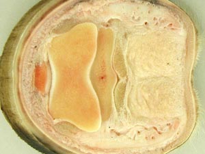
Horn-Producing Dermis
Lab Objectives:
• To identify the parts and regions of the equine hoof.
• To expose the collateral cartilages and digital cushion of the equine foot.
• To identify DIP and PIP joints and adjacent structures on a split equine foot.
• To identify the horn producing regions of the dermis on demonstration specimens.
• To identify the parts of the ruminant foot.
• To identify the parts of the porcine foot and be able to number the four digits.
Anatomical Terms:
Equine Foot Structure
coronet
periople
hoof
wall
toe
quarter
heel
bar
bulbs
collateral sulcus
sole
frog
apex of the frog
central sulcus of the frog
horny laminae
white line
coronary dermis
laminar dermis
collateral cartilages (of P3)
digital cushion
proximal interphalangeal (PIP) joint
distal interphalangeal (DIP) joint
navicular bursa
distal sesamoidean impar ligament
Ruminant & Camelid Foot Structures
claw
abaxial wall
axial wall
interdigital cleft
distal interdigital ligament
bulb
sole
coronet
dewclaw (digit 2 & 5, bov)
pedal joint (DIP)
pedal bone
Instructor Commentary:
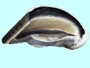
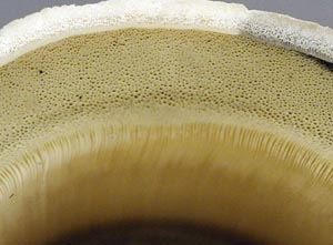
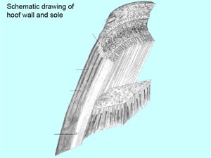
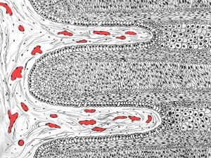
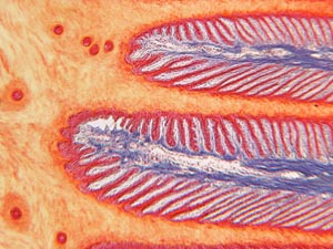
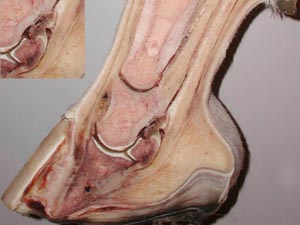
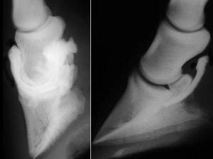
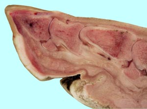
The foot is difficult to define but a common definition of the equine foot is the hoof and its contents. This leaves structures such as the collateral cartilages and P2 partly within and partly out of the hoof. The hoof is the capsule of the foot so the term "hoof capsule" is redundant. The hoof is horny tissue produced by specialized dermis. The hoof wall is produced by the coronary dermis and the laminar dermis produces the horny laminae on the inside of the hoof wall. Laminar units are formed by the interdigitation of horny insensitive laminae with adjacent sensitive dermal laminae. About 600 laminar units bear most or all of the body weight in the standing horse. Therefore, inflammation of the dermal laminae known as laminitis is very painful. Hoof horn is also produced by sole dermis and the frog dermis. An unshod horse on soft ground will bear some body weight on the sole and frog but the shod foot on a hard surface will bear all the weight on the laminae.
Hoof growth is less than a cm/month. Since the longest distance from the coronet to the ground surface is at the toe, the oldest, most dried out and least flexible part of the hoof is at the toe. In contrast, the heel is the most flexible part if the hoof wall. The sole is softer than the heel and the frog is the softest part of the hoof.
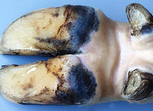
The ruminant foot is similar to the horse but it lacks the frog and collateral cartilages. The bulb of the heel in the equine foot does not contact the ground but in ruminants the bulb has ground contact and is weight bearing. Therefore, the laminae are not as well developed in ruminants and laminitis is less painful. The ruminant hoof wall is asymmetric with a flat axial wall and a curved abaxial wall. The axial wall is less well developed and therefore infection of the DIP joint occurs at the axial skin/wall junction.
Each ruminant foot has two hooves referred to as claws, an unfortunate term since they are quite different from the claws of carnivores. The vestigial digits are called dew claws because they are close enough to the ground to become wet from dew on grass. Pronghorn antelopes, giraffes and camelids lack dew claws but they are well developed in pigs. Like ruminants, the major digits of swine are 3 and 4 but digit 4 is larger than 3. The dewclaws of pigs are often elongated but this is not common in domestic ruminants. The bulb of ruminant feet is hard but it is soft in pigs. The hoof is well developed in pigs but the sole is small while the bulb is large and soft. Therefore, the pig foot is transitional between the soft padded foot of dogs and the hard hoof of ruminants.
Dissection Images:
Note: Click an image to see it enlarged, view its caption, and toggle its labels.
| 1 | 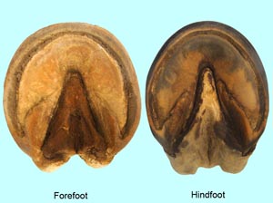 |
 |
2 |
| 3 | 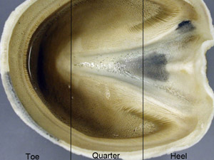 |
 |
4 |
| 5 | 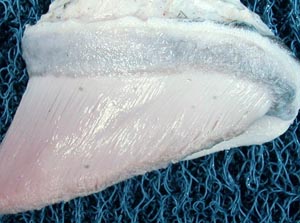 |
 |
6 |
| 7 | 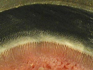 |
 |
8 |
| 9 | 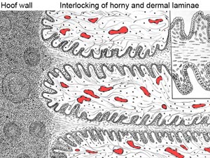 |
 |
10 |
| 11 | 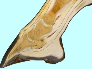 |
 |
12 |
| 13 | 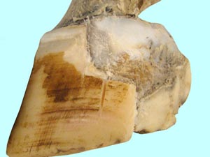 |
 |
14 |
| 15 | 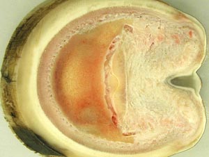 |
 |
16 |
| 17 | 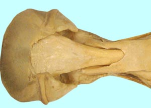 |
 |
18 |
| 19 | 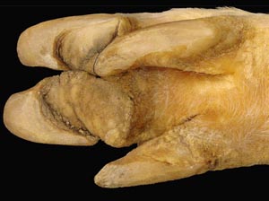 |
 |
20 |