
LAB 14 More Abdominal Viscera
Ruminant, Camelid, Pig, & Rabbit Viscera
Lab Objectives:
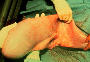
• Expose and palpate the abdominal viscera through paralumbar incisions in the abdominal wall.
• Through an incision in the caudal part of the dorsal ruminal sac on the left side, remove most of the ingesta and then palpate internal structures.
• Enlarge the right paralumbar incision and observe the layers of the omentum.
• After thorough palpation, remove the abdominal viscera as a single unit.
• Trace the path of ingesta from rumen to rectum.
• Examine the liver of ruminants and swine.
• Examine the umbilical vessels and abdominal viscera of a newborn calf.
• Examine permanent specimens of camelid stomachs and spiral colon.
• Examine the abdominal viscera of a rabbit and make comparisons with other herbivores.
Anatomical Terms:
Ruminant Abdominal Structures
paralumbar fossa
(term sequence follows demo viscera)
esophagus
spleen
greater omentum
superficial layer of the omentum
deep layer of the omentum
caudal fold of the omentum
rumen
papillae
dorsal, ventral and cranial sacs
dorsal & ventral caudal blind sacs
cranial & caudal pillars
ruminoreticular fold
reticulum
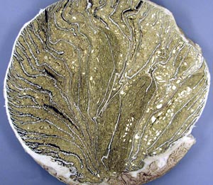
omasum
omasal laminae
abomasum
lesser omentum
pylorus
pyloric sphincter
torus pyloricus
caudal vena cava
liver
portal v.
hepatic a.
gallbladder
cystic duct
bile duct
hepatic veins
right, left & caudate lobes
falciform ligament
(duodenum)
descending duodenum
caudal flexure of the duodenum
jejunum
mesojejunum
mesenteric lymph nodes
ileum
ileocecal fold
ileal (ileocolic) orifice
cecum
(colon)
ascending colon
proximal loop of ascending colon
spiral colon
centripedal coils
central flexure
centrifugal coils
distal loop of ascending colon
bovine kidney
renal cortex
medulla
calyx
ureter
Swine Gastrointestinal Structures
liver
(stomach)
gastric diverticulum
torus pyloricus
jejunum
ileum
ileal papilla
cecum
colon
centripedal loop
central flexure
centrifugal loop
Instructor Commentary:
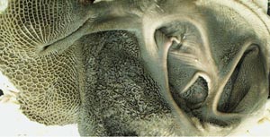
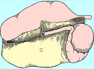
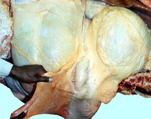
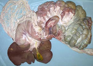
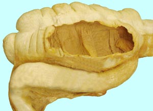
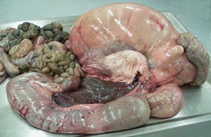
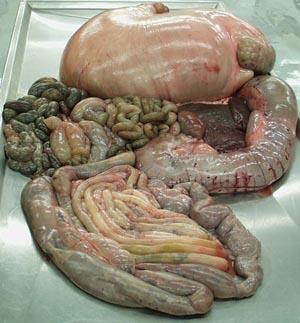
The ruminant abdomen is dominated by the voluminous ruminoreticulum which occupies the entire left side of the abdominal cavity. The reticulum is small when compared with the rumen but together they form a functional unit. The reticulum is lined by hexagonal cells while the rumen is lined with finger like papillae. Horizontal cranial and caudal pillars divide the rumen into dorsal, ventral and cranial sacs. Externally the pillars correspond to grooves with similar names so there is a caudal pillar and a caudal groove. The liver, omasum and abdomasum lie to the right of the cranial part of the ruminoreticulum. The liver which is most dorsal lies between the diaphragm and the omasum. The omasum is spherical and much firmer than other stomach compartments. It is positioned dorsal to the abomasum, a soft spindle shaped organ that lies near the midline and may be displaced to the left by passing under the cranial sac of the rumen.
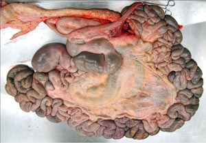
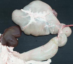
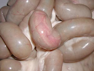
Ruminant intestines lie caudal to the abomasum on the right side of the rumen. The spiral colon is a two dimensional coil that is suspended in the sheet like mesocolon that covers the spiral colon on its right side. Therefore, the spiral colon is easily observed on the left side of the mesocolon but it is obscured on its right side by the mesocolon and the mesojejunum which is fused to the mesocolon so that the jejunum forms many loops around the periphery of the spiral colon. The intestines are surrounded by an omentum that is attached to the rumen on the left and on the right it attaches to the descending duodenum and the abomasum. The ruminant omentum is thicker and tougher than that of other species. It is so strong that is used for surgical correction of a displaced abdomen. This is done by suturing part of omentum to the body wall so that the closely attached abomasum is fixed and cannot displace
In some ways the gut of camelids resembles that of ruminants but there are significant differences and therefore camelids should not be referred to as ruminants. The camelid stomach has 3 compartments known as C1, C2, and C3. C1 is large like the rumen but lacks papillae and has glandular saccules. C2 is small like the reticulum but has glandular saccules that are more complex than those of C1. C3 is tubular like the abomasum but the glandular lining is significantly different. The spiral colon of camelids is a 2D coil as in ruminants but it is separate from the small intestine. The omentum of camelids is poorly developed.
The porcine stomach is small and simple like that of the horse and the lining has glandular and non-glandular parts. Also, like the horse, the porcine large intestine has bands and sacculations but they are more subtle in swine. Like ruminants and camelids, pigs have a spiral colon but it is three dimensional in pigs. The ileum enters the large intestine via an ileal papilla. Peyer's patches, first discovered in pig intestine, are organized lymphoid follicles that serve as inductive sites for mucosal immunity in the proximal jejunum. In the distal jejunum and ileum, Peyer's patches may serve instead as a first responder site capable of mounting humoral immune responses to pathogenic bacteria in the absence of T lymphocyte help.
The rabbit has a simple stomach and a large intestine with bands and sacculations. Most notable about the rabbit is a well developed cecal appendix that is rich in lymphoid tissue.
Dissection Images:
Note: Click an image to see it enlarged, view its caption, and toggle its labels.
| 1 | 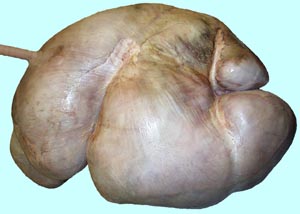 |
 |
2 |
| 3 | 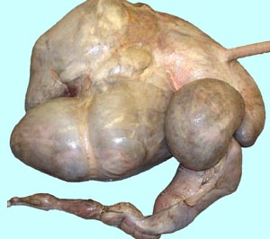 |
 |
4 |
| 5 | 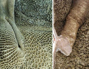 |
 |
6 |
| 7 | 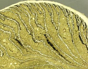 |
 |
8 |
| 9 | 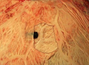 |
 |
10 |
| 11 | 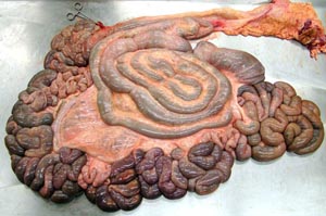 |
 |
12 |
| 13 | 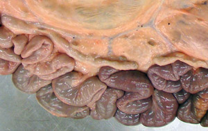 |
 |
14 |
| 15 | 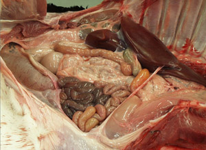 |
 |
16 |
| 17 | 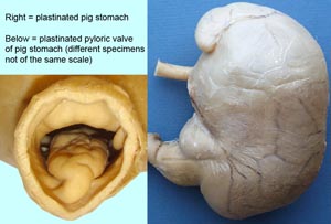 |
 |
18 |
| 19 | 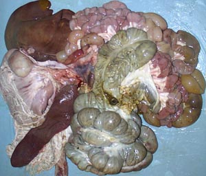 |
 |
20 |
| 21 | 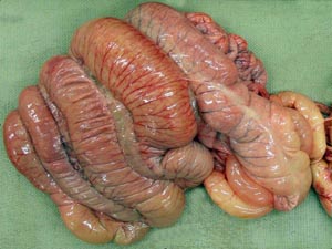 |
 |
22 |
| 23 | 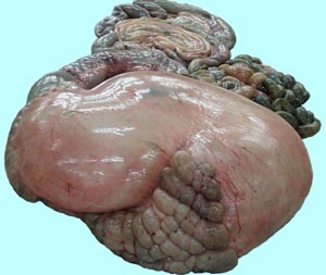 |
 |
24 |
| 25 | 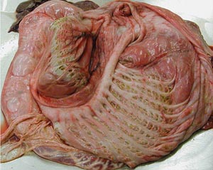 |
 |
26 |
| 27 | 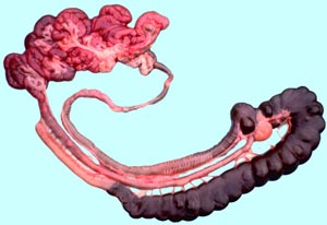 |
 |
28 |