Lab 16 - Image 2
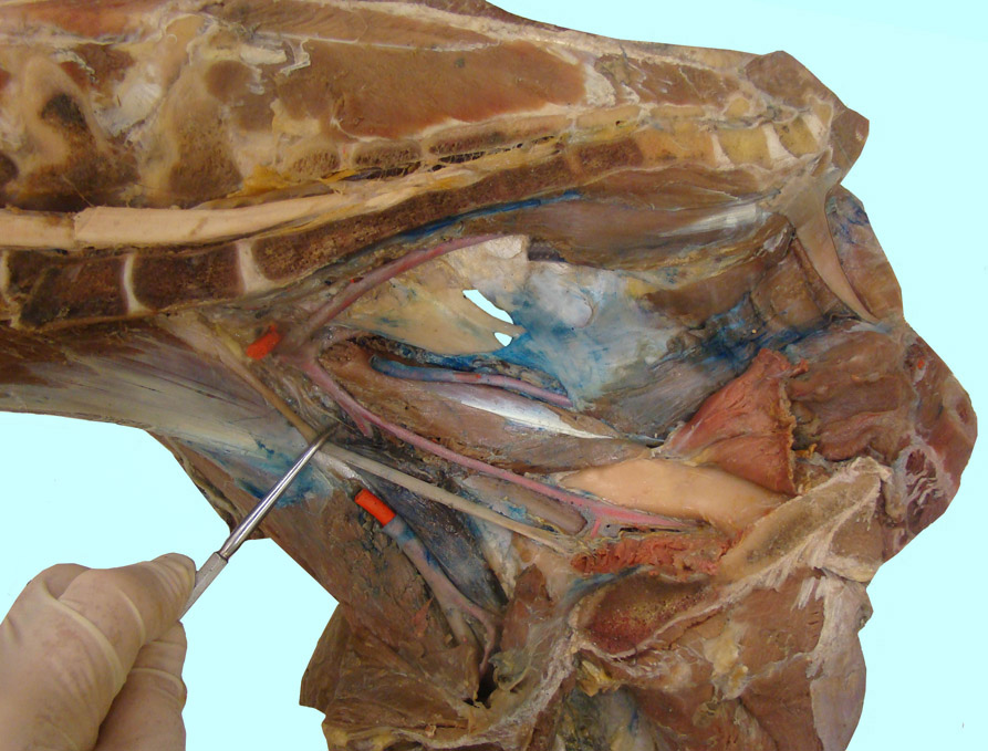
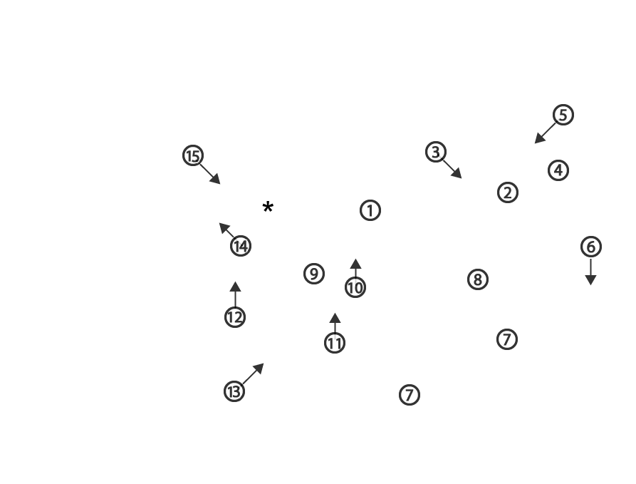
Equine pelvic dissection after removal of the pelvic viscera. The probe is pushing the obturator n. cranially. 1, sacrosciatic ligament; 2, levator ani m.; 3, coccygeus m.; 4, external anal sphincter m.; 5, retractor penis m.; 6, crus of the penis (sagittal section); 7, pelvic symphysis; 8, ventral part of the internal obturator m. reflected caudally; 9, dorsal part of internal obturator m.; 10, internal pudendal a.; 11, obturator a.; 12, iliacofemoral a.; 13, external iliac a.; 14, cranial gluteal a.; 15, caudal gluteal a.; asterisk, sciatic nerve.