Lab 18 - Image 5
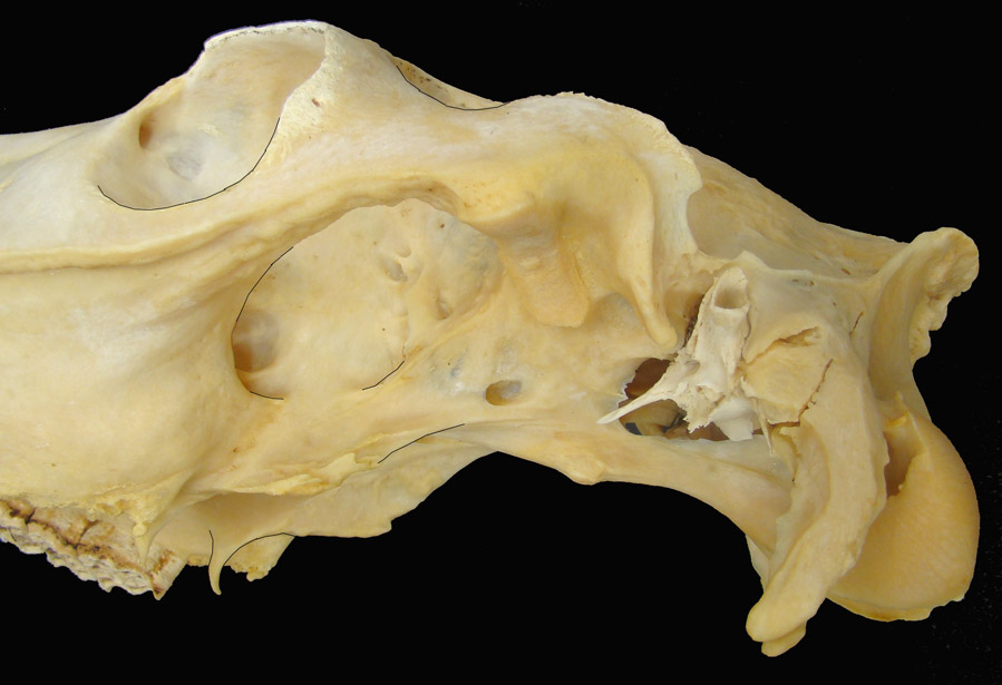
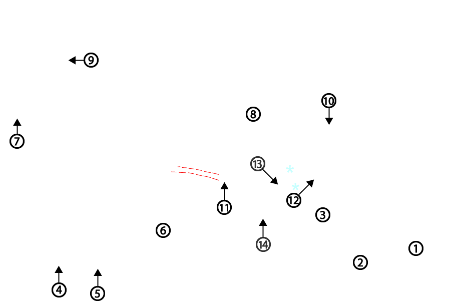
Lateral view of equine skull, caudal half. 1, occipital condyle; 2, paracondylar process; 3, basioccipital bone; 4, last molar tooth; 5, hamulus of the pterygoid bone; 6, vertical part of the palatine bone; 7, facial crest; 8, articular surface of the temporomandibular joint ; 9, entrance to the lacrimal canal; 10, external acoustic meatus; 11, caudal alar foramen; 12, attachment site for the stylohyoid bone (temporohyoid joint); 13, muscular process; 14, tuberosity for attachment of the longus capitus m.; asterisk, foramen lacerum; dashed red lines, location of the alar canal (the maxillary artery passes through here).