Lab 20 - Image 2
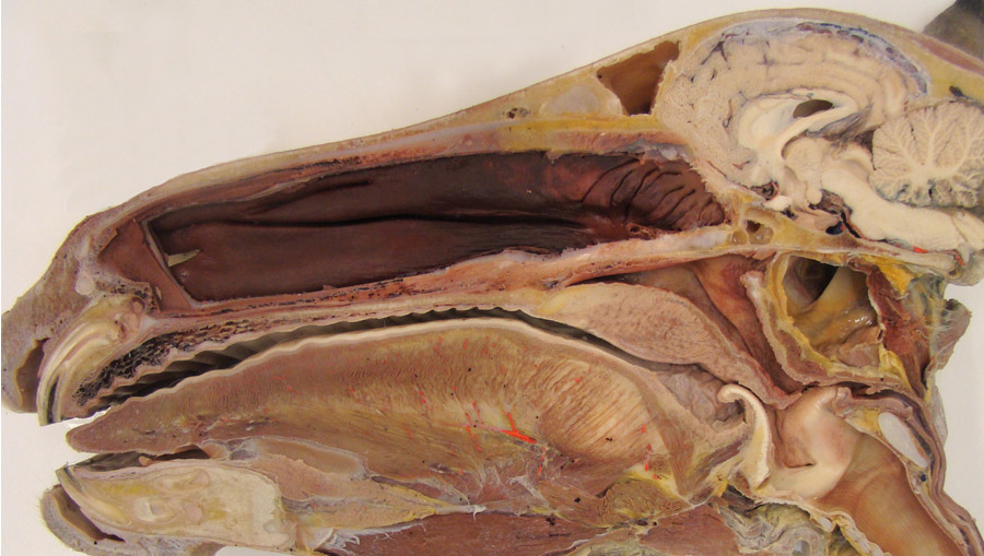

Equine split head after removal of the nasal septum to expose the nasal cavity. 1, dorsal concha; 2, ventral concha; 3 ethmoidal conchas; 4, vomer (bone); 5, frontal sinus; 6, hard palate; 7, soft palate; 8, orifice of the auditory tube on the lateral wall of the nasopharynx. At this place, an endoscope can be passed into the guttural pouch. 9, stylohyoid bone; 10, medial retropharyngeal lymph nodes adjacent to the ventral wall of the guttural pouch; 11, cricoid cartilage; 12, cricoid cartilage (ventral), 13, trachea; 14, ossified rostral edge of the thyroid cartilage; 15, basihyoid bone; asterisk, palatine tonsil.
NOTE: In this image the tip of the epiglottis is abnormally positioned ventral to the soft palate. The normal position is dorsal to the soft palate.
NOTE: In this image the tip of the epiglottis is abnormally positioned ventral to the soft palate. The normal position is dorsal to the soft palate.