
LAB 18 Introduction
Abdominal Vessels and
Pelvic Diaphragm & Nerves
(Guide to the Dissection of the Dog, 8th ed., pp. 174-184)
CONTENTS:
Lab Objectives:
• Identify branches of the abdominal aorta:
- paired arteries to the abdominal wall (lumbar arteries & deep circumflex iliac a.)
- paired arteries to viscera (renal a. & ovarian/testicular a.)
- unpaired arteries to viscera (celiac a., cranial mesenteric a., & caudal mesenteric a.)
• Examine the portal vein and its branches.
• Open the pelvic canal (by cutting through the pelvic symphysis) and find:
- the pelvic diaphragm (levator ani and coccygeus muscles)
- pelvic n. and pelvic plexus (including hypogastric n. input to the plexus)
- compartments of the peritoneal cavity
Anatomical Terms:
Branches of the Abdominal Aorta
lumbar arteries
celiac a.
hepatic a.
cystic a.
right gastric a.
gastroduodenal a.
right gastroepiploic a.
cranial pancreaticoduodenal a.
left gastric a.
esophageal branches
splenic a.
left gastroepiploic a. (aa.)
pancreatic branches
cranial mesenteric artery
common trunk (in the dog; usually separate branches in the cat)
middle colic a.
right colic a.
ileocolic a.
mesenteric ileal branch
colic branch
cecal a.
antimesenteric ileal branch
caudal pancreaticoduodenal a.
jejunal aa.
ileal aa.
phrenicoabdominal a. (common trunk)
cranial abdominal a.
caudal phrenic a.
renal aa. (right & left)
ovarian aa. (in mesovarium) / testicular aa. (in mesorchium) (right & left)
caudal mesenteric a.
left colic a.
cranial rectal a.
deep circumflex iliac a.
Portal Vein
gastroduodenal vein
splenic vein
left gastric vein
cranial & caudal mesenteric veins
Pelvic diaphragm & nerves
pelvic diaphragm:
levator ani m.
coccygeus m.
pelvic nerve & pelvic plexus
(right & left)
Extensions (excavations) of peritoneal cavity:
pararectal fossa
rectogenital pouch
vesicogenital pouch
pubovesical pouch
Instructor Commentary:
In general, body regions are supplied by arterial loops fed by more than one major artery. This arrangement is particularly obvious in the case of abdominal viscera because vessels can be viewed in mesenteries. Notice that branches of the celiac and cranial and caudal mesenteric arteries are connected through distal loops. Also, the abdominal aorta participates in top-level loops connecting the parent arteries.
The presence of vascular loops allows surgeons to ligate individual vessels with the expectation that blood will find its way to a particular region by alternate branches. However, the brain and the heart represent two regions where arterial looping is not robust. As a result, blockage of an artery in these regions often results in tissue death, leading to heart attacks and strokes.
All of the paired arteries that originate from the abdominal aorta have satellite veins that drain directly or indirectly into the caudal vena cava. However, blood conveyed by the three unpaired arteries supplying digestive organs and spleen must drain into the portal vein. The portal vein empties into liver sinusoids, from which blood reaches the caudal vena cava via hepatic veins.
As you prepare to identify the individual abdominal arteries that supply digestive organs and the spleen, keep the following overview in mind:
• The celiac a. supplies cranial viscera (spleen, liver, pancreas, esophagus, stomach, and descending duodenum). The caudal mesenteric a. supplies the descending colon and rectum. The cranial mesenteric a. supplies everything in between (ascending duodenum, jejunum, ileum, cecum, ascending and transverse colon).
• The celiac a. sends two branches to the left side (splenic and left gastric) and one to the right side (hepatic). The stomach receives a quadrant blood supply: left/right gastric arteries (lesser curvature) and left/right gastroepiploic arteries (greater curvature).
• The cranial mesenteric a. sends:
- a branch to the ascending duodenum (caudal pancreaticoduodenal a.),
- a common trunk (in the dog) to the large intestines:
ileocolic a. to the ileum (antimesenteric ileal a.), cecum, and adjacent colon;
right colic a. to ascending colon;
middle colic a. to transverse colon
- numerous jejunal arteries before terminating in a mesenteric ileal artery
Dissection Steps:
Click to view a PDF list of dissection procedures for this lab:
Show List of Dissection Steps (PDF)
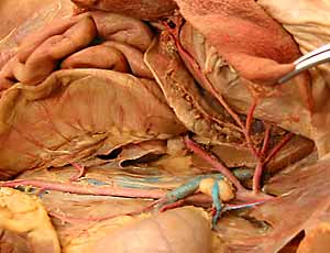
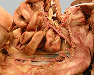
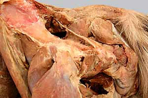
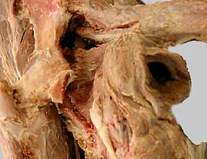
Dissection Images:
Note: Click an image to see it enlarged, view its caption, and toggle its labels.
| 1 | 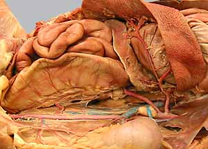 |
 |
2 |
| 3 | 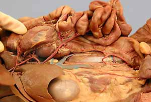 |
 |
4 |
| 5 | 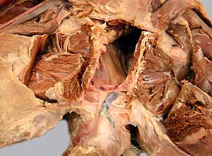 |
 |
6 |
| 7 | 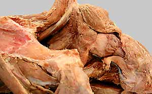 |
 |
8 |