
LAB 25 Introduction
Superficial Nerves, Head Arteries,
and Cervical Structures
(Guide to the Dissection of the Dog, 8th ed., pp. 260-276)
CONTENTS:
Lab Objectives:
• Dissect superficial nerves of the head:
- facial nerve and its branches (innervate facial expression muscles)
- mandibular nerve branches: lingual nerve & inferior alveolar nerve
also, as you dissect vessels, find:
- maxillary nerve, which becomes infraorbital n. (p. 259)
- vagus nerve and its laryngeal branches (p. 260)
- hypoglossal nerve (p. 260)
• Examine certain structures located at the neck-head juncture:
- thyroid gland and external parathyroid gland
- esophagus
- tracheal cartilages and tracheal smooth muscle
- medial retropharyngeal lymph node
• Identify branches of the common carotid artery:
- internal carotid artery (dog) & carotid sinus (located on the occipital a. in the cat)
- external carotid artery, and its major branches:
-- occipital artery
-- lingual artery
-- facial artery
-- maxillary artery (continues the external carotid artery), and its major branches:
--- inferior alveolar artery
--- external ophthalmic artery
--- infraorbital artery (continuation of the maxillary artery)
Anatomical Terms:
Head: nerves
facial nerve (cranial nerve VII)
caudal auricular branches
ventral buccal n.
dorsal buccal n.
auriculopalpebral n.
rostral auricular branches
palpebral branches
mandibular nerve (a branch of V, trigeminal nerve)
auriculotemporal n.
buccal n.
lingual n.
inferior alveolar n.
mylohyoid n.
maxillary nerve (a branch of V, trigeminal nerve)
infraorbital n.
vagus nerve (cranial nerve X)
cranial laryngeal n.
caudal laryngeal n. (termination of the recurrent laryngeal n.)
cervical sympathetic trunk
cranial cervical ganglion
hypoglossal nerve (cranial nerve XII)
Cervical Structures:
thyroid gland [palpate]
external & internal parathyroid glands
esophagus
pharyngoesophageal limen
trachea [palpate]
tracheal cartilages
medial retropharyngeal lymph node [palpate]
Head: arteries
common carotid artery [palpate pulse]
caudal & cranial thyroid aa.
internal carotid artery (proximal portion absent in adult cats)
carotid sinus
external carotid artery
occipital artery
ascending pharyngeal artery (assumes internal carotid a. function in adult cats)
cranial laryngeal artery
lingual artery
facial a.
sublingual a.
caudal auricular a.
superficial temporal a.
maxillary a. (continues external carotid a.)
inferior alveolar a.
caudal deep temporal a.
middle meningeal a.
external ophthalmic a.
palatine aa.
infraorbital a. (continues maxillary a.)
Note:
limen [Latin = threshold; beginning boundary] e.g., pharyngoesophageal limen
alveolus [Latin = small hollow] = tooth socket (e.g., inferior alveolar n.)
occipital [Latin: occiput = back of the head] e.g., occipital a.
ophthalmic [Greek: ophthalmos = eye] e.g., external ophthalmic a.
Instructor Commentary:
Even though it looks like two (bilateral) glands, the carnivore thyroid gland is regarded as a single gland with bilateral lobes because it originates embryologically as a single bi-lobed mass from the ventral surface of the pharynx (small contributions come from the fourth pharyngeal pouch). The thyroid gland remains single in some species (pig). In other species (horse), the bilateral lobes are connected typically by an isthmus. In the carnivore, the bilateral lobes are infrequently connected by an isthmus.
You may have first hand experience with regional nerve blocks that you received prior to dentistry procedures. The lower jaw is anesthetized by depositing local anesthetic on the inferior alveolar nerve just before it enters the mandibular canal. Upper cheek teeth are anesthetized by blocking the maxillary nerve before it enters the infraorbital canal. Upper incisor teeth can be blocked by depositing anesthetic on branches of the infraorbital nerve.
Dissection Steps:
Click to view a PDF list of dissection procedures for this lab:
Show List of Dissection Steps (PDF)
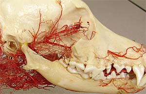
Dissection Images:
Note: Click an image to see it enlarged, view its caption, and toggle its labels.
| 1 | 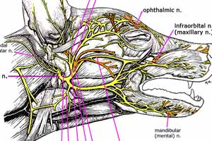 |
 |
2 |
| 3 |  |
 |
4 |
| 5 |  |
 |
6 |
| 7 |  |
 |
8 |
| 9 |  |
 |
10 |
| 11 |  |
 |
12 |
| 13 | 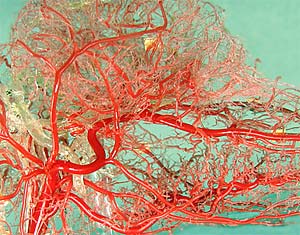 |
 |
14 |